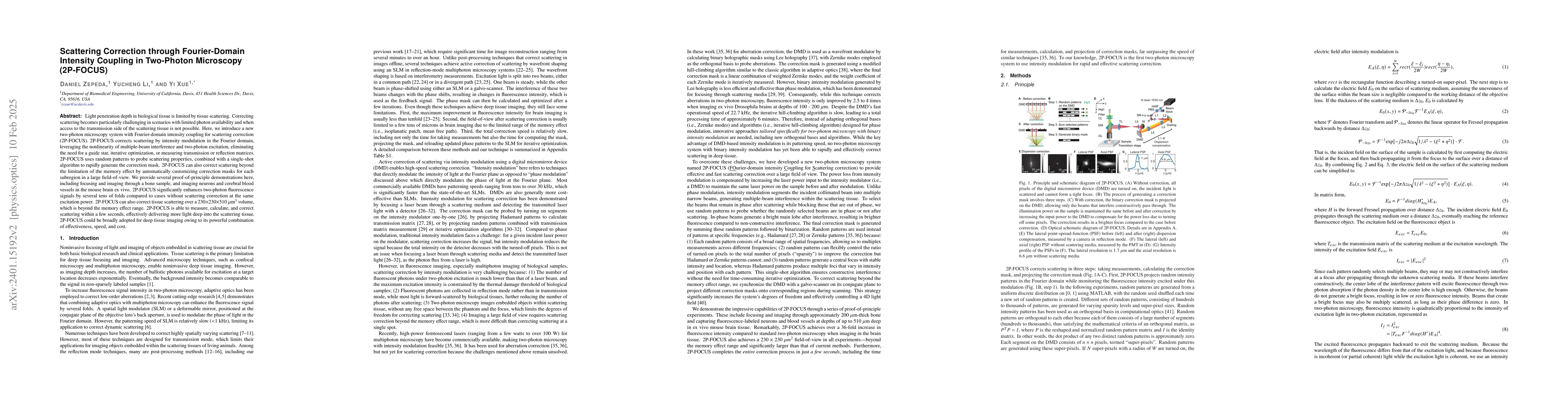
Scattering compensation through Fourier-domain open-channel coupling in two-photon microscopy
Publication
Metrics
AI Quick Summary
This paper introduces 2P-FOCUS, a two-photon microscopy system that compensates for scattering in biological tissue using Fourier-domain open-channel coupling, significantly enhancing two-photon fluorescence signals without requiring guide stars or matrix measurements. The method enables deep tissue imaging in live animals by correcting scattering over large volumes.
Paper Preview
Abstract
Light penetration depth in biological tissue is limited by tissue scattering. There is an urgent need for scattering compensation in vivo focusing and imaging, particularly challenging in photon-starved scenarios, without access to the transmission side of the scattering tissue. Here, we introduce a two-photon microscopy system with Fourier-domain open-channel coupling for scattering correction (2P-FOCUS). 2P-FOCUS corrects scattering by utilizing the non-linearity of multiple-beam interference and two-photon excitation, eliminating the need for a guide star, iterative optimization, or measuring transmission or reflection matrices. We demonstrate that 2P-FOCUS significantly enhances two-photon fluorescence signals by several tens of folds when focusing through a bone sample, compared to cases without scattering compensation at equivalent excitation power. We also show that 2P-FOCUS can correct scattering over large volumes by imaging neurons and cerebral blood vessels within a 230x230x500 um\textsuperscript{3} volume in the mouse brain in vitro. 2P-FOCUS could serve as a powerful tool for deep tissue imaging in bulky organisms or live animals.
AI Key Findings
Get AI-generated insights about this paper's methodology, results, significance, and more — seven facets brought into focus.
Impact
Paper Details
Authors
PDF Preview
Key Terms
Citation Network
Current paper (gray), citations (green), references (blue)
Display is limited for performance on very large graphs.
Discussion 0