ArxivLens

Quick Summary:

The paper presents SD-LayerNet, a semi-supervised retinal layer segmentation method for OCT images that leverages anatomical priors and disentangled representations. This approach effectively utilizes both labeled and unlabeled data, improving segmentation performance even with limited labeled data.

Journal Reference: MICCAI 2022. Lecture Notes in Computer Science, vol 13438. Springer, Cham
DOI: 10.1007/978-3-031-16452-1_31
arXiv Id: 2207.00458
Comments: Accepted at MICCAI 2022
Date Published: 2022-10-26
Date Updated: 2022-10-26

Categories:

Key Terms (TF-IDF):

segmentation (0.237)
segmentation (0.228)
priors (0.203)
priors (0.198)
layer (0.193)
layer (0.186)
pixel (0.184)
pixel (0.184)
labeled (0.174)
labeled (0.173)
supervised (0.150)
supervised (0.150)
state art (0.146)
art (0.145)
state art (0.144)
art (0.143)
factors (0.142)
representations (0.140)
factors (0.137)
representations (0.135)
License: http://creativecommons.org/licenses/by/4.0/

Summary:

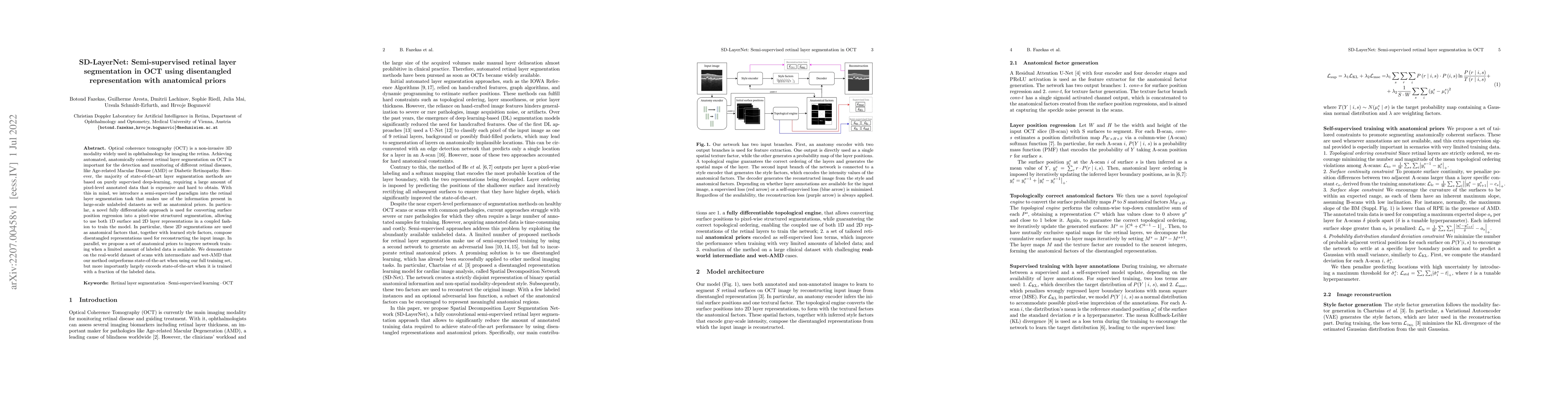
Optical coherence tomography (OCT) is a non-invasive 3D modality widely used in ophthalmology for imaging the retina. Achieving automated, anatomically coherent retinal layer segmentation on OCT is important for the detection and monitoring of different retinal diseases, like Age-related Macular Disease (AMD) or Diabetic Retinopathy. However, the majority of state-of-the-art layer segmentation methods are based on purely supervised deep-learning, requiring a large amount of pixel-level annotated data that is expensive and hard to obtain. With this in mind, we introduce a semi-supervised paradigm into the retinal layer segmentation task that makes use of the information present in large-scale unlabeled datasets as well as anatomical priors. In particular, a novel fully differentiable approach is used for converting surface position regression into a pixel-wise structured segmentation, allowing to use both 1D surface and 2D layer representations in a coupled fashion to train the model. In particular, these 2D segmentations are used as anatomical factors that, together with learned style factors, compose disentangled representations used for reconstructing the input image. In parallel, we propose a set of anatomical priors to improve network training when a limited amount of labeled data is available. We demonstrate on the real-world dataset of scans with intermediate and wet-AMD that our method outperforms state-of-the-art when using our full training set, but more importantly largely exceeds state-of-the-art when it is trained with a fraction of the labeled data.

Citation & Reference Network Visualization

This graph shows how this paper is connected to other research. The current paper is shown in dark gray, papers that cite it are in green, and papers it references are in blue. You can:

Current Paper
Papers citing this work (Citations)
Papers cited by this work (References)

• Hover over any node to see the paper title

• Click a node to view that paper's details

• Drag nodes to rearrange the graph

• Use the zoom controls (bottom right) to zoom in/out

Citations and References

Title Authors Year Actions
Wide-field OCT volumetric segmentation using semi-supervised CNN and transformer integration
Syna Sreng, Padmini Ramesh, Pham Duc Nam Phuong, Nur Fidyana Binte Abdul Gani, Jacqueline Chua, M. Nongpiur, Tin Aung, R. Husain, Leopold Schmetterer, D. Wong
2025
View Paper
SD-LayerNet: Robust and label-efficient retinal layer segmentation via anatomical priors
Botond Fazekas, Guilherme Aresta, Dmitry A. Lachinov, Sophie Riedl, Julia Mai, U. Schmidt-Erfurth, H. Bogunović
2025
View Paper
Formula-Driven Data Augmentation and Partial Retinal Layer Copying for Retinal Layer Segmentation
Tsubasa Konno, Takahiro Ninomiya, Kanta Miura, Koichi Ito, Noriko Himori, Parmanand Sharma, Toru Nakazawa, T. Aoki
2024
View Paper
Detecting Retinopathy From Optical Coherence Tomography Images Using a Novel Augmentation-Based Semi-Supervised Learning Approach
Bowen Zheng, Chenxi Huang, Yuan Li, Zhiyuan Zheng, Yuemei Luo
2024
View Paper
Deep Neural Networks for Automated Outer Plexiform Layer Subsidence Detection on Retinal OCT of Patients With Intermediate AMD
Guilherme Aresta, Teresa Araújo, G. Reiter, Julia Mai, Sophie Riedl, C. Grechenig, Robyn H Guymer, Zhichao Wu, U. Schmidt-Erfurth, H. Bogunović
2024
View Paper
Deep learning based multimodal biomedical data fusion: An overview and comparative review
J. Duan, Jiaqi Xiong, Yinghui Li, Weiping Ding
2024
View Paper
Point based weakly semi-supervised biomarker detection with cross-scale and label assignment in retinal OCT images
Xiaoming Liu, Xing-Li Zhu, Ying Zhang, Man Wang
2024
View Paper
Diverse Data Generation for Retinal Layer Segmentation With Potential Structure Modeling
Kun Huang, Xiao Ma, Zetian Zhang, Yuhan Zhang, Songtao Yuan, Huazhu Fu, Qiang Chen
2024
View Paper
Semi-supervised information fusion for medical image analysis: Recent progress and future perspectives
Ying Weng, Yiming Zhang, Wenxin Wang, Tom Dening
2024
View Paper
Weakly Semi-supervised object detection with point annotations in Retinal OCT images
Xiaoming Liu, Xing-Li Zhu, Jinshan Tang
2023
View Paper
TranSegNet: Hybrid CNN-Vision Transformers Encoder for Retina Segmentation of Optical Coherence Tomography
Yiheng Zhang, Zhongliang Li, Nan Nan, Xiangzhao Wang
2023
View Paper

Similar Papers

Uncertainty-aware retinal layer segmentation in OCT through probabilistic signed distance functions

Erik J Bekkers, Clara I. Sánchez, Bart Liefers, Caroline Klaver, Mohammad Mohaiminul Islam, Coen de Vente

Published: 2024-12-06

Retinal OCT Synthesis with Denoising Diffusion Probabilistic Models for Layer Segmentation

Peter Walter, Dennis Eschweiler, Johannes Stegmaier, Yuli Wu, Weidong He, Ningxin Dou, Zixin Fan, Shengli Mi

Published: 2024-03-08

Comments (0)

This website uses cookies to ensure you get the best experience. Learn more