Publication
Metrics
AI Quick Summary
This paper presents a piezoresponse force microscopy technique for visualizing moiré superlattices in van der Waals heterostructures with sub-5 nm resolution, demonstrating flexoelectric effects in diverse materials, including graphene and transition metal dichalcoogenides. The method reveals that moiré superlattices function as networks of polarized domain walls.
Paper Preview
Abstract
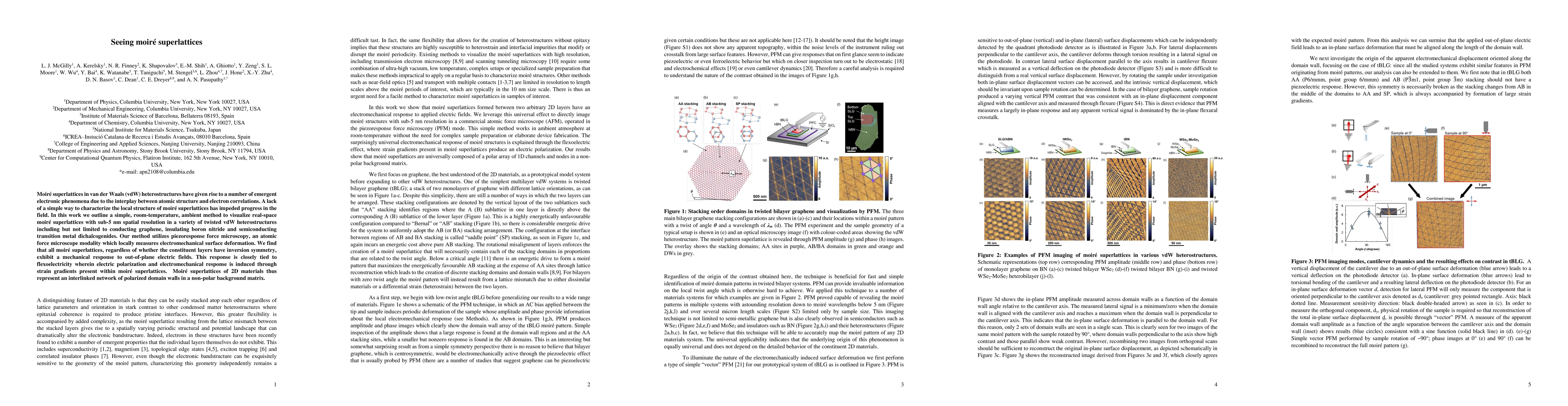
Moir\'e superlattices in van der Waals (vdW) heterostructures have given rise to a number of emergent electronic phenomena due to the interplay between atomic structure and electron correlations. A lack of a simple way to characterize moir\'e superlattices has impeded progress in the field. In this work we outline a simple, room-temperature, ambient method to visualize real-space moir\'e superlattices with sub-5 nm spatial resolution in a variety of twisted vdW heterostructures including but not limited to conducting graphene, insulating boron nitride and semiconducting transition metal dichalcogenides. Our method utilizes piezoresponse force microscopy, an atomic force microscope modality which locally measures electromechanical surface deformation. We find that all moir\'e superlattices, regardless of whether the constituent layers have inversion symmetry, exhibit a mechanical response to out-of-plane electric fields. This response is closely tied to flexoelectricity wherein electric polarization and electromechanical response is induced through strain gradients present within moir\'e superlattices. Moir\'e superlattices of 2D materials thus represent an interlinked network of polarized domain walls in a non-polar background matrix.
AI Key Findings
Get AI-generated insights about this paper's methodology, results, significance, and more — seven facets brought into focus.
Impact
Paper Details
PDF Preview
Key Terms
Citation Network
Current paper (gray), citations (green), references (blue)
Display is limited for performance on very large graphs.
Discussion 0