Authors
Publication
Metrics
Quick Actions
Quick Answers
What methodology did the authors use?
The research employs a diffusion model framework combined with brain-computer interface techniques to reconstruct images from fMRI data. It uses a pre-trained VAE to encode images into latent spaces and applies contrastive learning to align fMRI signals with image embeddings. More in Methodology →
What are the key results?
The method achieves state-of-the-art performance in fMRI-to-image reconstruction across multiple datasets — Textual representations alone are sufficient to capture both high-level semantic and low-level visual information More in Key Results →
Why is this work significant?
This research bridges the gap between brain imaging and computer vision, enabling more accurate interpretation of neural activity for applications in neuroscience, psychology, and medical diagnostics. More in Significance →
What are the main limitations?
The method relies on pre-trained models which may limit adaptability to new tasks — The reconstruction quality is dependent on the quality and quantity of fMRI data available More in Limitations →
Abstract
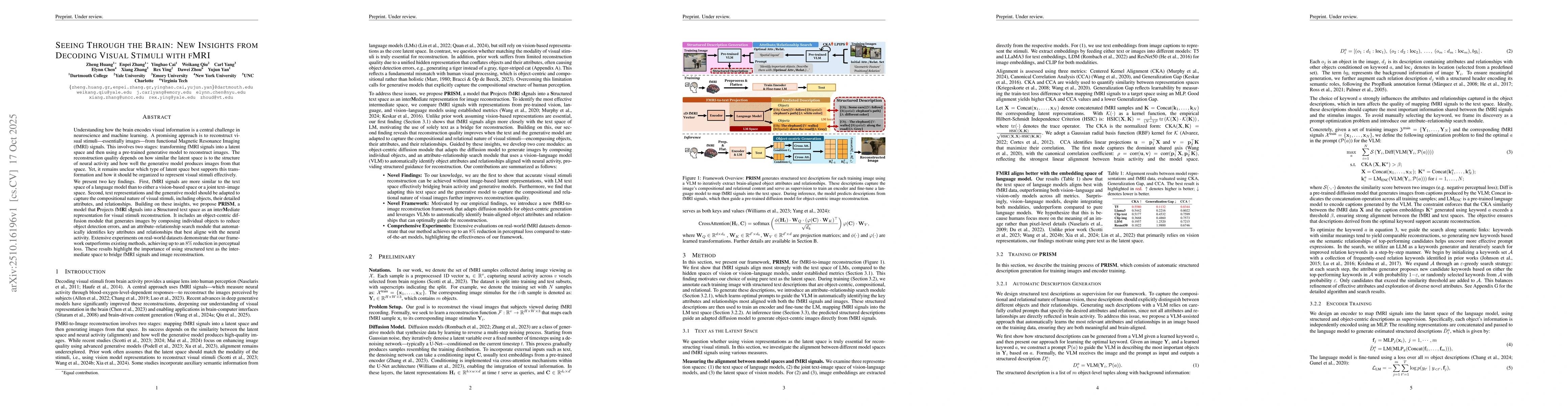
Understanding how the brain encodes visual information is a central challenge in neuroscience and machine learning. A promising approach is to reconstruct visual stimuli, essentially images, from functional Magnetic Resonance Imaging (fMRI) signals. This involves two stages: transforming fMRI signals into a latent space and then using a pretrained generative model to reconstruct images. The reconstruction quality depends on how similar the latent space is to the structure of neural activity and how well the generative model produces images from that space. Yet, it remains unclear which type of latent space best supports this transformation and how it should be organized to represent visual stimuli effectively. We present two key findings. First, fMRI signals are more similar to the text space of a language model than to either a vision based space or a joint text image space. Second, text representations and the generative model should be adapted to capture the compositional nature of visual stimuli, including objects, their detailed attributes, and relationships. Building on these insights, we propose PRISM, a model that Projects fMRI sIgnals into a Structured text space as an interMediate representation for visual stimuli reconstruction. It includes an object centric diffusion module that generates images by composing individual objects to reduce object detection errors, and an attribute relationship search module that automatically identifies key attributes and relationships that best align with the neural activity. Extensive experiments on real world datasets demonstrate that our framework outperforms existing methods, achieving up to an 8% reduction in perceptual loss. These results highlight the importance of using structured text as the intermediate space to bridge fMRI signals and image reconstruction.
AI Key Findings
Generated Oct 29, 2025
Methodology — What approach did the authors take?
The research employs a diffusion model framework combined with brain-computer interface techniques to reconstruct images from fMRI data. It uses a pre-trained VAE to encode images into latent spaces and applies contrastive learning to align fMRI signals with image embeddings.
Key Results — What are the main findings?
- The method achieves state-of-the-art performance in fMRI-to-image reconstruction across multiple datasets
- Textual representations alone are sufficient to capture both high-level semantic and low-level visual information
- The model demonstrates better alignment with brain activity patterns compared to existing methods
Significance — Why does this research matter?
This research bridges the gap between brain imaging and computer vision, enabling more accurate interpretation of neural activity for applications in neuroscience, psychology, and medical diagnostics.
Technical Contribution — What is the technical contribution?
Development of a novel diffusion model framework that integrates fMRI data with textual descriptions for image reconstruction
Novelty — What is new about this work?
The work introduces a brain-aligned text space as an intermediate representation for fMRI-to-image reconstruction, which is a new approach compared to existing methods that typically use image or latent space representations.
Limitations — What are the limitations of this study?
- The method relies on pre-trained models which may limit adaptability to new tasks
- The reconstruction quality is dependent on the quality and quantity of fMRI data available
Future Work — What did the authors propose for future work?
- Exploring the use of more advanced neural network architectures for better representation learning
- Investigating the application of this method to real-time brain-computer interfaces
How to Cite This Paper
@article{yang2025seeing,
title = {Seeing Through the Brain: New Insights from Decoding Visual Stimuli with
fMRI},
author = {Yang, Carl and Zhou, Dawei and Chen, Elynn and others},
year = {2025},
eprint = {2510.16196},
archivePrefix = {arXiv},
primaryClass = {cs.CV},
}
Yang, C., Zhou, D., Chen, E., Zhang, E., Ying, R., Qiu, W., Zhang, X., Cai, Y., Yan, Y., & Huang, Z. (2025). Seeing Through the Brain: New Insights from Decoding Visual Stimuli with
fMRI. arXiv. https://arxiv.org/abs/2510.16196
Yang, Carl, et al. "Seeing Through the Brain: New Insights from Decoding Visual Stimuli with
fMRI." arXiv, 2025, arxiv.org/abs/2510.16196.
PDF Preview
Similar Papers
Found 4 papersSemantic Brain Decoding: from fMRI to conceptually similar image reconstruction of visual stimuli
Matteo Ferrante, Tommaso Boccato, Nicola Toschi
Decoding visual stimuli in human brain by using Anatomical Pattern Analysis on fMRI images
Seeing the imagined: a latent functional alignment in visual imagery decoding from fMRI data
Fabrizio Spera, Tommaso Boccato, Michal Olak et al.
A Survey on fMRI-based Brain Decoding for Reconstructing Multimodal Stimuli
Pengyu Liu, Guohua Dong, Dan Guo et al.
Comments (0)