Summary
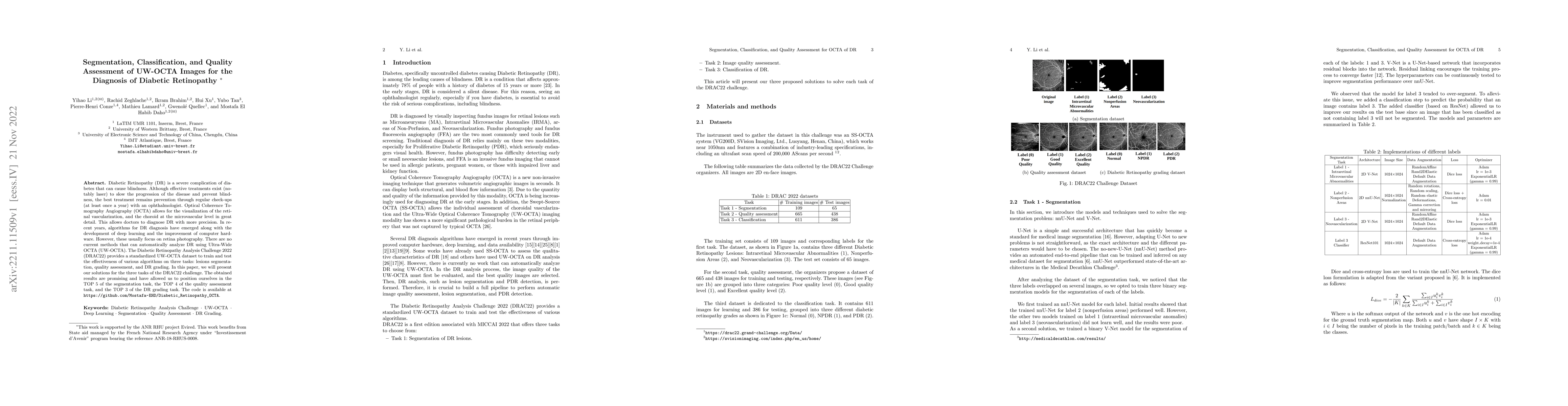
Diabetic Retinopathy (DR) is a severe complication of diabetes that can cause blindness. Although effective treatments exist (notably laser) to slow the progression of the disease and prevent blindness, the best treatment remains prevention through regular check-ups (at least once a year) with an ophthalmologist. Optical Coherence Tomography Angiography (OCTA) allows for the visualization of the retinal vascularization, and the choroid at the microvascular level in great detail. This allows doctors to diagnose DR with more precision. In recent years, algorithms for DR diagnosis have emerged along with the development of deep learning and the improvement of computer hardware. However, these usually focus on retina photography. There are no current methods that can automatically analyze DR using Ultra-Wide OCTA (UW-OCTA). The Diabetic Retinopathy Analysis Challenge 2022 (DRAC22) provides a standardized UW-OCTA dataset to train and test the effectiveness of various algorithms on three tasks: lesions segmentation, quality assessment, and DR grading. In this paper, we will present our solutions for the three tasks of the DRAC22 challenge. The obtained results are promising and have allowed us to position ourselves in the TOP 5 of the segmentation task, the TOP 4 of the quality assessment task, and the TOP 3 of the DR grading task. The code is available at \url{https://github.com/Mostafa-EHD/Diabetic_Retinopathy_OCTA}.
AI Key Findings
Get AI-generated insights about this paper's methodology, results, and significance.
Paper Details
PDF Preview
Key Terms
Citation Network
Current paper (gray), citations (green), references (blue)
Display is limited for performance on very large graphs.
Similar Papers
Found 4 papersSemi-Supervised Semantic Segmentation Methods for UW-OCTA Diabetic Retinopathy Grade Assessment
Zeyu Ding, Zhuoyi Tan, Hizmawati Madzin
Deep-OCTA: Ensemble Deep Learning Approaches for Diabetic Retinopathy Analysis on OCTA Images
Jilan Xu, Rui Feng, Yuejie Zhang et al.
Improved Automatic Diabetic Retinopathy Severity Classification Using Deep Multimodal Fusion of UWF-CFP and OCTA Images
Yihao Li, Mostafa El Habib Daho, Pierre-Henri Conze et al.
DRAC: Diabetic Retinopathy Analysis Challenge with Ultra-Wide Optical Coherence Tomography Angiography Images
Hao Chen, Jaeyoung Kim, Bin Sheng et al.
| Title | Authors | Year | Actions |
|---|
Comments (0)