ArxivLens

Quick Summary:

This paper introduces a novel segmentation framework to detect early-stage pancreatic neuroendocrine tumors (PNETs) from abdominal CT scans, utilizing a new dataset of 376 cases. The method achieves high sensitivity and specificity, outperforming existing segmentation networks, indicating potential for clinical impact in early cancer detection.

arXiv Id: 2004.02021
Date Published: 2020-04-07
Date Updated: 2020-04-07

Categories:

Key Terms (TF-IDF):

cancer (0.297)
ct (0.290)
cancer (0.263)
checking (0.239)
tumor (0.234)
checking (0.225)
quantitatively (0.219)
segmentation (0.207)
best knowledge (0.204)
knowledge (0.201)
quantitatively (0.195)
cases (0.180)
segmentation (0.179)
knowledge (0.175)
best (0.170)
cases (0.160)
dataset (0.157)
task (0.152)
specificity (0.151)
best (0.143)
License: http://arxiv.org/licenses/nonexclusive-distrib/1.0/

Summary:

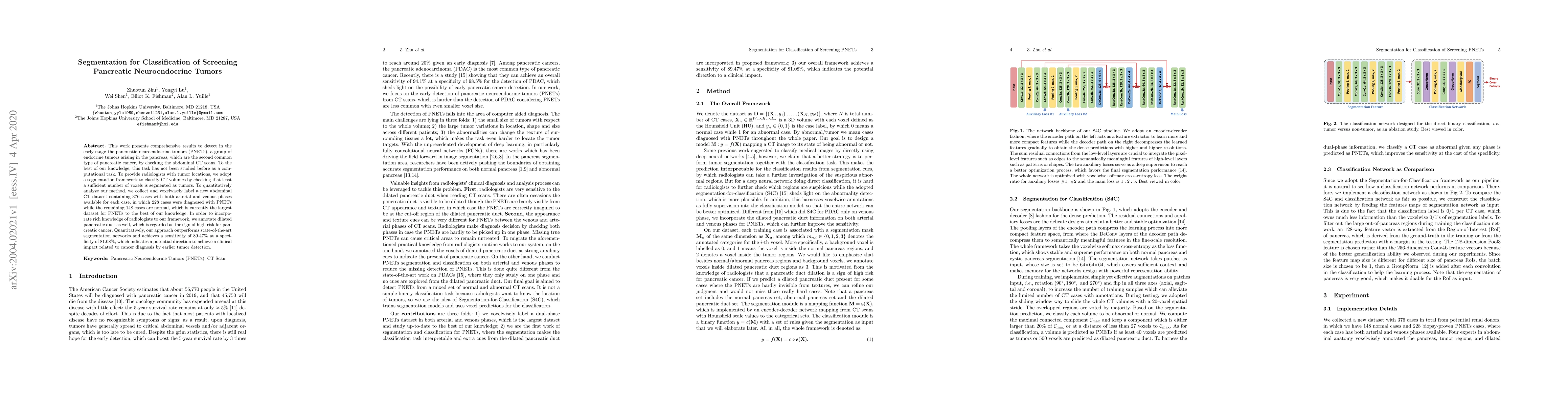
This work presents comprehensive results to detect in the early stage the pancreatic neuroendocrine tumors (PNETs), a group of endocrine tumors arising in the pancreas, which are the second common type of pancreatic cancer, by checking the abdominal CT scans. To the best of our knowledge, this task has not been studied before as a computational task. To provide radiologists with tumor locations, we adopt a segmentation framework to classify CT volumes by checking if at least a sufficient number of voxels is segmented as tumors. To quantitatively analyze our method, we collect and voxelwisely label a new abdominal CT dataset containing $376$ cases with both arterial and venous phases available for each case, in which $228$ cases were diagnosed with PNETs while the remaining $148$ cases are normal, which is currently the largest dataset for PNETs to the best of our knowledge. In order to incorporate rich knowledge of radiologists to our framework, we annotate dilated pancreatic duct as well, which is regarded as the sign of high risk for pancreatic cancer. Quantitatively, our approach outperforms state-of-the-art segmentation networks and achieves a sensitivity of $89.47\%$ at a specificity of $81.08\%$, which indicates a potential direction to achieve a clinical impact related to cancer diagnosis by earlier tumor detection.

Citation & Reference Network Visualization

This graph shows how this paper is connected to other research. The current paper is shown in dark gray, papers that cite it are in green, and papers it references are in blue. You can:

Current Paper
Papers citing this work (Citations)
Papers cited by this work (References)

• Hover over any node to see the paper title

• Click a node to view that paper's details

• Drag nodes to rearrange the graph

• Use the zoom controls (bottom right) to zoom in/out

Citations and References

Title Authors Year Actions
Neuroendocrine Tumor Detection System
Mohd Umair Kidwai, Anuradha Misha, Atul Srivastava, Vijay Shankar Sharma
2024
View Paper
Improved Pancreatic Cancer Detection and Localization on CT Scans: A Computer-Aided Detection Model Utilizing Secondary Features
M. Ramaekers, Christiaan G. A. Viviers, Terese A. E. Hellström, Lotte J S Ewals, Nick Tasios, Igor Jacobs, Joost Nederend, F. V. D. Sommen, M. Luyer
2024
View Paper
Segmentation-based Assessment of Tumor-Vessel Involvement for Surgical Resectability Prediction of Pancreatic Ductal Adenocarcinoma
Christiaan G. A. Viviers, M. Ramaekers, Amaan Valiuddin, Terese A. E. Hellström, Nick Tasios, John C. P. van der Ven, Igor Jacobs, Lotte J S Ewals, Joost Nederend, Peter H N de With, M. Luyer, F. van der Sommen
2023
View Paper
Automatic Detection of Pancreatic Lesions and Main Pancreatic Duct Dilatation on Portal Venous CT Scans Using Deep Learning
Clément Abi Nader, Rebeca Vétil, Laura Kate Wood, Marc-Michel Rohé, Alexandre Bône, H. Karteszi, M. Vullierme
2023
View Paper
Meta-information-aware Dual-path Transformer for Differential Diagnosis of Multi-type Pancreatic Lesions in Multi-phase CT
Bo Zhou, Yingda Xia, Jiawen Yao, Le Lu, Jingren Zhou, Chi Liu, J. Duncan, Ling Zhang
2023
View Paper
CT classification model of pancreatic serous cystic neoplasm and mucinous cystic neoplasm based on deep transfer learning.
Jin Li, Wei Yin, Yuanjun Wang
2022
View Paper
Integrating features from lymph node stations for metastatic lymph node detection
Chaoyi Wu, Feng Chang, Xiao Su, Zhihan Wu, Yanfeng Wang, Ling Zhu, Ya Zhang
2022
View Paper
3D Graph Anatomy Geometry-Integrated Network for Pancreatic Mass Segmentation, Diagnosis, and Quantitative Patient Management
Tianyi Zhao, Kai Cao, Jiawen Yao, Isabella Nogues, Le Lu, Lingyun Huang, Jing Xiao, Zhaozheng Yin, Ling Zhang
2020
View Paper
Lymph Node Gross Tumor Volume Detection and Segmentation via Distance-based Gating using 3D CT/PET Imaging in Radiotherapy
Zhuotun Zhu, D. Jin, K. Yan, T. Ho, X. Ye, Dazhou Guo, Chun-Hung Chao, Jing Xiao, A. Yuille, Le Lu
2020
View Paper
Pancreatic Cancer Imaging: A New Look at an Old Problem.
L. Chu, Seyoun Park, S. Kawamoto, A. Yuille, R. Hruban, E. Fishman
2020
View Paper

Similar Papers

An Exceptional Dataset For Rare Pancreatic Tumor Segmentation

Xinpeng Zhang, Zhenxing Qian, Yue Yan, Wei Tang, Xiaoxiao Hu, Wenqi Li, Keyang Zhou, Yingli Chen, Zilu Zheng

Published: 2025-01-29

Optimizing Synthetic Data for Enhanced Pancreatic Tumor Segmentation

Linkai Peng, Zheyuan Zhang, Gorkem Durak, Frank H. Miller, Alpay Medetalibeyoglu, Michael B. Wallace, Ulas Bagci

Published: 2024-07-27

A Weakly Supervised Segmentation Network Embedding Cross-scale Attention Guidance and Noise-sensitive Constraint for Detecting Tertiary Lymphoid Structures of Pancreatic Tumors

Jun Chen, Liang Mao, Xiaoping Yang, Zhongqiu Wang, Yingying Cao, Jingya Chen, Bingxue Wang, Liwen Zou, Zhenghua Cai, Yudong Qiu, Luying Gui

Published: 2023-07-28

Comments (0)

This website uses cookies to ensure you get the best experience. Learn more