Segmentation of Brain Metastases in MRI: A Two-Stage Deep Learning Approach with Modality Impact Study
Publication
Metrics
AI Quick Summary
This study develops a two-stage deep learning model for segmenting brain metastases in MRI, demonstrating that combining T1c, T1, and FLAIR modalities yields better results than single-modality or all-modalities approaches. The proposed model sets a new benchmark in accuracy, emphasizing the significance of modality selection and multi-stage processing.
Paper Preview
Abstract
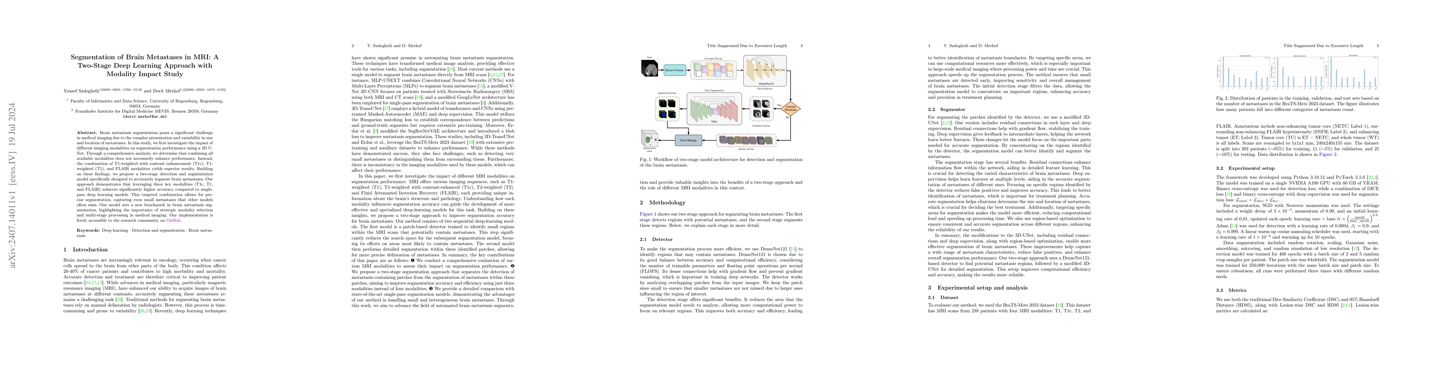
Brain metastasis segmentation poses a significant challenge in medical imaging due to the complex presentation and variability in size and location of metastases. In this study, we first investigate the impact of different imaging modalities on segmentation performance using a 3D U-Net. Through a comprehensive analysis, we determine that combining all available modalities does not necessarily enhance performance. Instead, the combination of T1-weighted with contrast enhancement (T1c), T1-weighted (T1), and FLAIR modalities yields superior results. Building on these findings, we propose a two-stage detection and segmentation model specifically designed to accurately segment brain metastases. Our approach demonstrates that leveraging three key modalities (T1c, T1, and FLAIR) achieves significantly higher accuracy compared to single-pass deep learning models. This targeted combination allows for precise segmentation, capturing even small metastases that other models often miss. Our model sets a new benchmark in brain metastasis segmentation, highlighting the importance of strategic modality selection and multi-stage processing in medical imaging. Our implementation is freely accessible to the research community on \href{https://github.com/xmindflow/Met-Seg}{GitHub}.
AI Key Findings
Get AI-generated insights about this paper's methodology, results, significance, and more — seven facets brought into focus.
Impact
Authors
PDF Preview
Citation Network
Current paper (gray), citations (green), references (blue)
Display is limited for performance on very large graphs.
Discussion 0