Segmentation of Cortical Spreading Depression Wavefronts Through Local Similarity Metric
Publication
Metrics
AI Quick Summary
This paper introduces a new segmentation method for cortical spreading depressions using local intensity similarity metrics in 2-photon microscopy images, achieving a DICE index of 0.9859 and a 79.9% reduction in root mean square error, outperforming current state-of-the-art techniques.
Paper Preview
Abstract
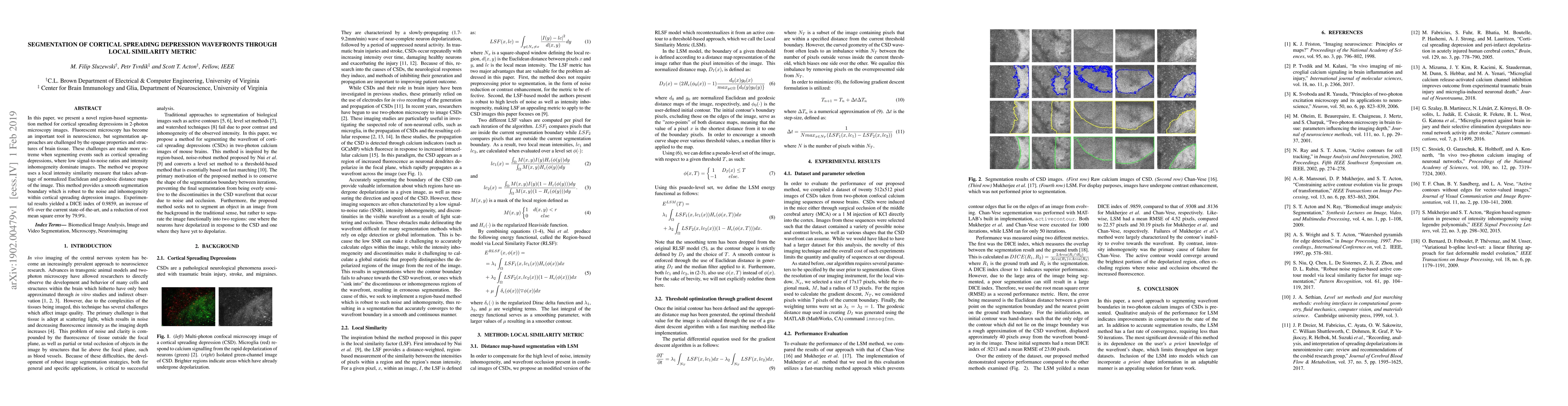
In this paper, we present a novel region-based segmentation method for cortical spreading depressions in 2-photon microscopy images. Fluorescent microscopy has become an important tool in neuroscience, but segmentation approaches are challenged by the opaque properties and structures of brain tissue. These challenges are made more extreme when segmenting events such as cortical spreading depressions, where low signal-to-noise ratios and intensity inhomogeneity dominate images. The method we propose uses a local intensity similarity measure that takes advantage of normalized Euclidean and geodesic distance maps of the image. This method provides a smooth segmentation boundary which is robust to the noise and inhomogeneity within cortical spreading depression images. Experimental results yielded a DICE index of 0.9859, an increase of 6% over the current state-of-the-art, and a reduction of root mean square error by 79.9%.
AI Key Findings
Get AI-generated insights about this paper's methodology, results, significance, and more — seven facets brought into focus.
Impact
Paper Details
PDF Preview
Key Terms
Citation Network
Current paper (gray), citations (green), references (blue)
Display is limited for performance on very large graphs.
Discussion 0