Summary
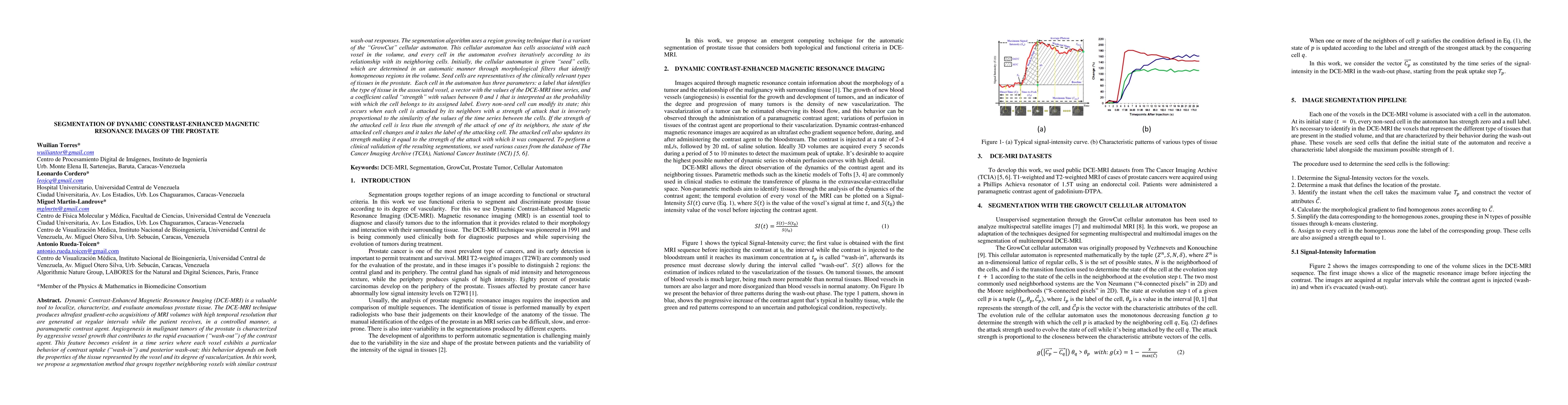
Dynamic Contrast-Enhanced Magnetic Resonance Imaging (DCE-MRI) is a valuable tool to localize, characterize, and evaluate anomalous prostate tissue. Ultrafast gradient-echo acquisitions of MRI volumes are generated at regular time intervals while the patient receives a paramagnetic contrast agent. The result is a time series where each voxel exhibits a particular behavior of contrast uptake (wash-in) and posterior wash-out. In this work, a segmentation method is proposed that groups together neighboring voxels with similar contrast wash-out responses, using a variant of the region growing GrowCut cellular automaton algorithm, that evolves iteratively according to the relationship of a cell to its neighboring cells. Initially, seed cells are determined through morphological filters that identify homogeneous regions in the volume that are representatives of the clinically relevant types of tissues in the prostate. Each cell is characterized by three parameters: a label that identifies the type of tissue in the associated voxel, a vector with the values of the DCE-MRI time series, and a strength coefficient with values between 0 and 1 that represents the probability with which the cell belongs to its assigned label. Every non-seed cell can modify its state; this occurs when each cell is attacked by its neighbors with a strength of attack that is inversely proportional to the similarity of the values of the time series between the cells. If the strength of the attacked cell is less than the strength of the attack of one of its neighbors, its state changes to the one of the attacking cell. The attacked cell also updates its strength making it equal to the strength of the attack with which it was conquered. To perform a clinical validation of the resulting segmentations, we used various cases from the database of The Cancer Imaging Archive (TCIA), National Cancer Institute (NCI).
AI Key Findings
Get AI-generated insights about this paper's methodology, results, and significance.
Paper Details
PDF Preview
Key Terms
Citation Network
Current paper (gray), citations (green), references (blue)
Display is limited for performance on very large graphs.
Similar Papers
Found 4 papersDiffusion Models for Contrast Harmonization of Magnetic Resonance Images
Matthias Weigel, Cristina Granziera, Robin Sandkühler et al.
No citations found for this paper.
Comments (0)