Quick Summary:

This paper proposes an automated method using a 3D fully convolutional neural network for segmenting intracranial carotid artery calcification (ICAC) to aid in stroke risk assessment. The method incorporates deep supervision, skip connections, and dropout regularization to improve segmentation accuracy, achieving a Dice score of 76.2% and high correlation with manual annotations.

Summary

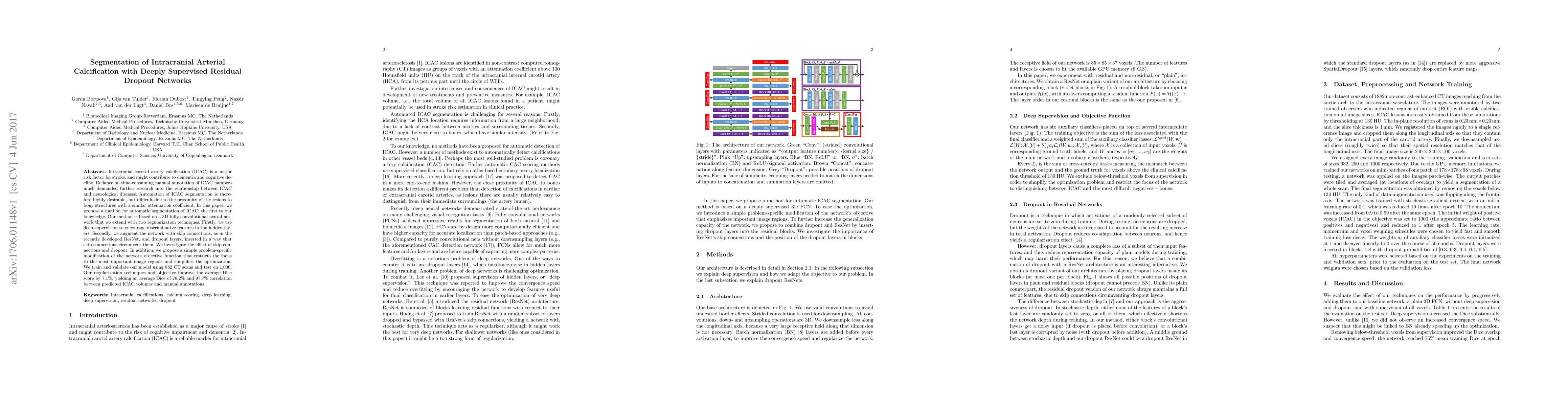
Intracranial carotid artery calcification (ICAC) is a major risk factor for stroke, and might contribute to dementia and cognitive decline. Reliance on time-consuming manual annotation of ICAC hampers much demanded further research into the relationship between ICAC and neurological diseases. Automation of ICAC segmentation is therefore highly desirable, but difficult due to the proximity of the lesions to bony structures with a similar attenuation coefficient. In this paper, we propose a method for automatic segmentation of ICAC; the first to our knowledge. Our method is based on a 3D fully convolutional neural network that we extend with two regularization techniques. Firstly, we use deep supervision (hidden layers supervision) to encourage discriminative features in the hidden layers. Secondly, we augment the network with skip connections, as in the recently developed ResNet, and dropout layers, inserted in a way that skip connections circumvent them. We investigate the effect of skip connections and dropout. In addition, we propose a simple problem-specific modification of the network objective function that restricts the focus to the most important image regions and simplifies the optimization. We train and validate our model using 882 CT scans and test on 1,000. Our regularization techniques and objective improve the average Dice score by 7.1%, yielding an average Dice of 76.2% and 97.7% correlation between predicted ICAC volumes and manual annotations.

AI Key Findings

Get AI-generated insights about this paper's methodology, results, and significance.

Paper Details

PDF Preview

Download PDF

Key Terms

connections (0.228)
manual (0.207)
layers (0.204)
supervision (0.203)
regularization (0.187)
hidden (0.182)
segmentation (0.180)
objective (0.157)
network (0.145)
average (0.142)
ct (0.139)
diseases (0.134)
techniques (0.133)
volumes (0.132)
annotations (0.129)
cognitive (0.128)
recently developed (0.128)
objective function (0.126)
model using (0.126)
proximity (0.125)

Citation Network

Current paper (gray), citations (green), references (blue)

Current Paper
Citations
References
Click to view
Title Authors Year Actions
Pre-processing and Quality Control of Large Clinical CT Head Datasets for Intracranial Arterial Calcification Segmentation
Benjamin Jin, Maria del C. Vald'es Hern'andez, Alessandro Fontanella, Wenwen Li, Eleanor Platt, Paul Armitage, A. Storkey, Joanna M. Wardlaw, Grant Mair
2024
View Paper
Calcified Tissue Detection using Deep Learning Models in Intravascular Ultrasound images
Priyanka Arora, Parminder Singh, A. Girdhar
2023
View Paper
Calcification Detection in Intravascular Ultrasound (IVUS) Images Using Transfer Learning Based MultiSVM model
Priyanka Arora, Parminder Singh, A. Girdhar, R. Vijayvergiya
2023
View Paper
Automated Detection of Cervical Carotid Artery Calcifications in Cone Beam Computed Tomographic Images Using Deep Convolutional Neural Networks
M. Ajami, Pavani Tripathi, H. Ling, M. Mahdian
2022
View Paper
A Comprehensive Review on Deep Supervision: Theories and Applications
Renjie Li, Xinyi Wang, Guangtai Huang, Wenli Yang, Kaining Zhang, Xiaodong Gu, S. Tran, Saurabh Garg, J. Alty, Quan Bai
2022
View Paper
MICDIR: Multi-scale Inverse-consistent Deformable Image Registration using UNetMSS with Self-Constructing Graph Latent
S. Chatterjee, Himanshi Bajaj, Istiyak H. Siddiquee, N. Subbarayappa, Steve Simon, S. Shashidhar, O. Speck, A. Nürnberger
2022
View Paper
Automated Segmentation and Volume Measurement of Intracranial Carotid Artery Calcification on Non-Contrast CT
G. Bortsova, D. Bos, Florian Dubost, M. Vernooij, M. Ikram, Gijs van Tulder, Marleen de Bruijne
2021
View Paper
DS6, Deformation-Aware Semi-Supervised Learning: Application to Small Vessel Segmentation with Noisy Training Data
S. Chatterjee, H. Mattern, Marleen de Bruijne
2020
View Paper
Regularized siamese neural network for unsupervised outlier detection on brain multiparametric magnetic resonance imaging: Application to epilepsy lesion screening
Zaruhi Alaverdyan, Julien Jung, R. Bouet, C. Lartizien
2019
View Paper
Medical Image Segmentation Using Deep Learning
Karen López-Linares Román, María Inmaculada García Ocaña, Nerea Lete Urzelai, M. Ballester, Iván Macía Oliver
2019
View Paper
Unsupervised representation learning for anomaly detection on neuroimaging. Application to epilepsy lesion detection on brain MRI. (Apprentissage de représentation non supervisé pour la détection d'anomalies en neuroimagerie. Application à la détection de lésions épileptogènes en Imagerie IRM)
Zaruhi Alaverdyan
2019
View Paper
Calcification Detection of Coronary Artery Disease in Intravascular Ultrasound Image: Deep Feature Learning Approach
Hannah Sofian, J. Than, Suraya Mohammad, N. Noor
2018
View Paper
Fully Convolutional Neural Networks for Road Detection with Multiple Cues Integration
Xiaofeng Han, Jianfeng Lu, Chunxia Zhao, Hongdong Li
2018
View Paper
3D regression neural network for the quantification of enlarged perivascular spaces in brain MRI
Florian Dubost, H. Adams, G. Bortsova, M. Ikram, W. Niessen, M. Vernooij, Marleen de Bruijne
2018
View Paper
GP-Unet: Lesion Detection from Weak Labels with a 3D Regression Network
Florian Dubost, G. Bortsova, H. Adams, M. Ikram, W. Niessen, M. Vernooij, Marleen de Bruijne
2017
View Paper

Comments (0)

This website uses cookies to ensure you get the best experience. Learn more