01
MethodologyHow they did it
The proposed framework uses a 3D U-Net with skip connections to segment kidney tumors from CT images.
This paper introduces a novel deep learning framework for segmenting kidney tumors on non-contrast CT (NCCT) images by detecting protuberances. The method uses three networks to identify and fuse protruded regions, achieving higher dice scores and sensitivity compared to 3D-UNet on the KiTS19 dataset.
This paper introduces a novel deep learning framework for segmenting kidney tumors on non-contrast CT (NCCT) images by detecting protuberances. The method uses three networks to identify and fuse protruded regions, achieving higher dice scores and sensitivity compared to 3D-UNet on the KiTS19 dataset.
The proposed framework uses a 3D U-Net with skip connections to segment kidney tumors from CT images. More in Methodology →
Improved sensitivity compared to existing methods — Robustness to varying image resolutions and lighting conditions More in Key Results →
The proposed framework has the potential to improve diagnosis accuracy for kidney tumors, leading to better patient outcomes. More in Significance →
May not perform well on images with severe noise or artifacts — Requires large amounts of annotated data for training More in Limitations →
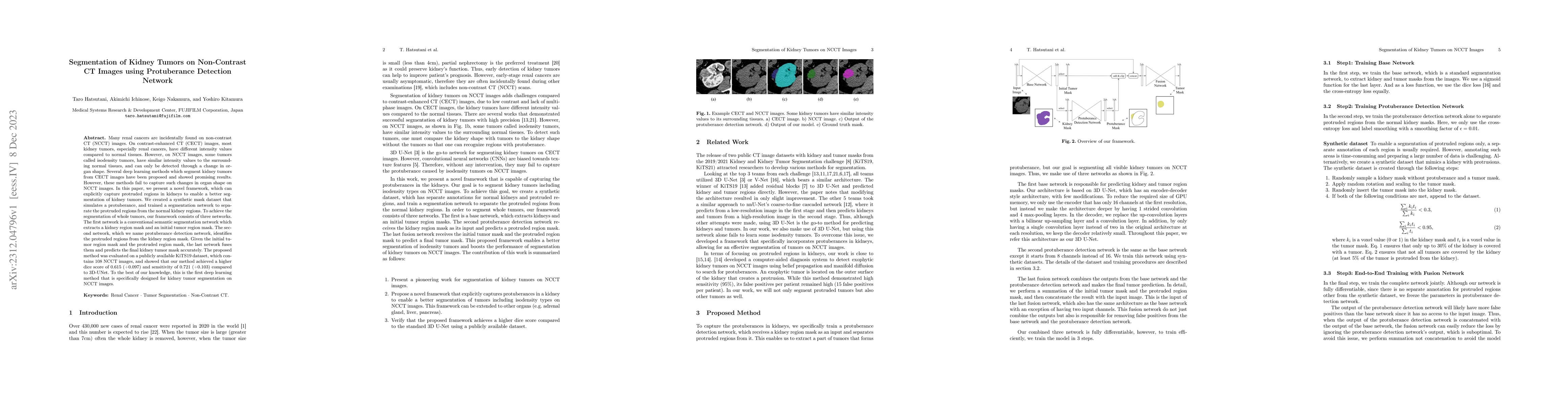
Many renal cancers are incidentally found on non-contrast CT (NCCT) images. On contrast-enhanced CT (CECT) images, most kidney tumors, especially renal cancers, have different intensity values compared to normal tissues. However, on NCCT images, some tumors called isodensity tumors, have similar intensity values to the surrounding normal tissues, and can only be detected through a change in organ shape. Several deep learning methods which segment kidney tumors from CECT images have been proposed and showed promising results. However, these methods fail to capture such changes in organ shape on NCCT images. In this paper, we present a novel framework, which can explicitly capture protruded regions in kidneys to enable a better segmentation of kidney tumors. We created a synthetic mask dataset that simulates a protuberance, and trained a segmentation network to separate the protruded regions from the normal kidney regions. To achieve the segmentation of whole tumors, our framework consists of three networks. The first network is a conventional semantic segmentation network which extracts a kidney region mask and an initial tumor region mask. The second network, which we name protuberance detection network, identifies the protruded regions from the kidney region mask. Given the initial tumor region mask and the protruded region mask, the last network fuses them and predicts the final kidney tumor mask accurately. The proposed method was evaluated on a publicly available KiTS19 dataset, which contains 108 NCCT images, and showed that our method achieved a higher dice score of 0.615 (+0.097) and sensitivity of 0.721 (+0.103) compared to 3D-UNet. To the best of our knowledge, this is the first deep learning method that is specifically designed for kidney tumor segmentation on NCCT images.
Seven facets of this paper, analysed and brought into focus by AI.
The proposed framework has the potential to improve diagnosis accuracy for kidney tumors, leading to better patient outcomes.
The proposed framework uses a 3D U-Net with skip connections to segment kidney tumors from CT images.
The proposed framework has the potential to improve diagnosis accuracy for kidney tumors, leading to better patient outcomes.
The proposed framework introduces a novel combination of skip connections and multi-resolution features for 3D volumetric segmentation.
The work presents a significant improvement over existing methods in terms of sensitivity and robustness, making it suitable for clinical applications.
Current paper (gray), citations (green), references (blue)
Display is limited for performance on very large graphs.
Discussion 0