Segmentation of MRI head anatomy using deep volumetric networks and multiple spatial priors
Publication
Metrics
AI Quick Summary
This paper proposes a Multiprior network that integrates spatial, morphological, and contextual priors into a deep convolutional network to improve MRI head anatomy segmentation, especially in the presence of lesions. The network outperforms existing segmentation tools and shows promising results in clinical applications for patients with disorders of consciousness.
Paper Preview
Abstract
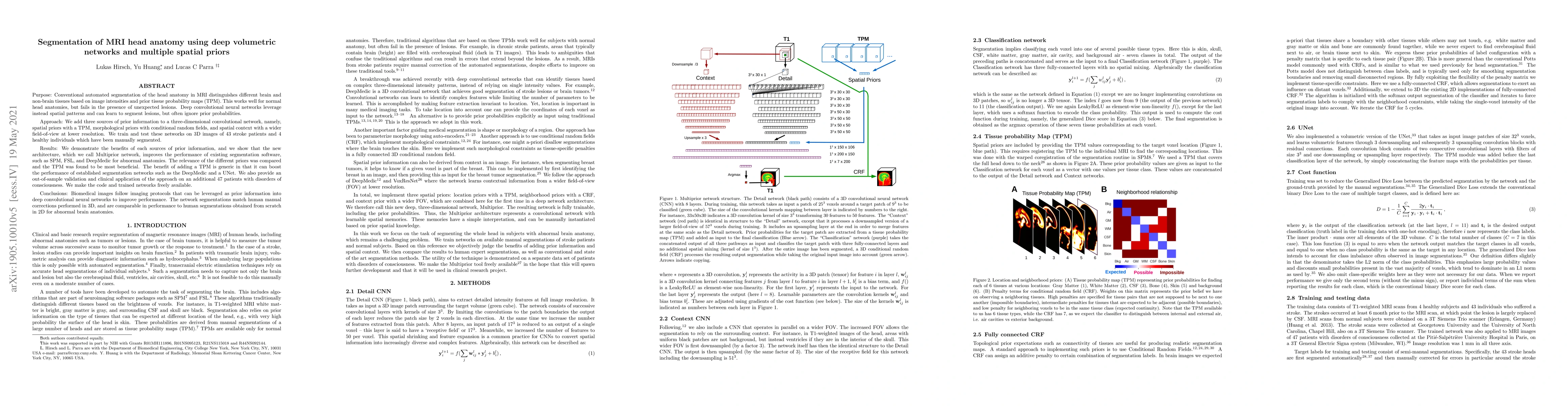
Purpose: Conventional automated segmentation of the head anatomy in MRI distinguishes different brain and non-brain tissues based on image intensities and prior tissue probability maps (TPM). This works well for normal head anatomies, but fails in the presence of unexpected lesions. Deep convolutional neural networks leverage instead spatial patterns and can learn to segment lesions, but often ignore prior probabilities. Approach: We add three sources of prior information to a three-dimensional convolutional network, namely, spatial priors with a TPM, morphological priors with conditional random fields, and spatial context with a wider field-of-view at lower resolution. We train and test these networks on 3D images of 43 stroke patients and 4 healthy individuals which have been manually segmented. Results: We demonstrate the benefits of each sources of prior information, and we show that the new architecture, which we call Multiprior network, improves the performance of existing segmentation software, such as SPM, FSL, and DeepMedic for abnormal anatomies. The relevance of the different priors was compared and the TPM was found to be most beneficial. The benefit of adding a TPM is generic in that it can boost the performance of established segmentation networks such as the DeepMedic and a UNet. We also provide an out-of-sample validation and clinical application of the approach on an additional 47 patients with disorders of consciousness. We make the code and trained networks freely available. Conclusions: Biomedical images follow imaging protocols that can be leveraged as prior information into deep convolutional neural networks to improve performance. The network segmentations match human manual corrections performed in 3D, and are comparable in performance to human segmentations obtained from scratch in 2D for abnormal brain anatomies.
AI Key Findings
Get AI-generated insights about this paper's methodology, results, significance, and more — seven facets brought into focus.
Impact
Paper Details
PDF Preview
Key Terms
Citation Network
Current paper (gray), citations (green), references (blue)
Display is limited for performance on very large graphs.
Discussion 0