This paper proposes a novel method for segmenting overlapped steatosis regions in high-resolution liver histopathology images, utilizing bounding box estimation and high-curvature point detection for precise segmentation. Experimental results indicate the method's potential to improve steatosis quantification in liver disease pathology reviews.
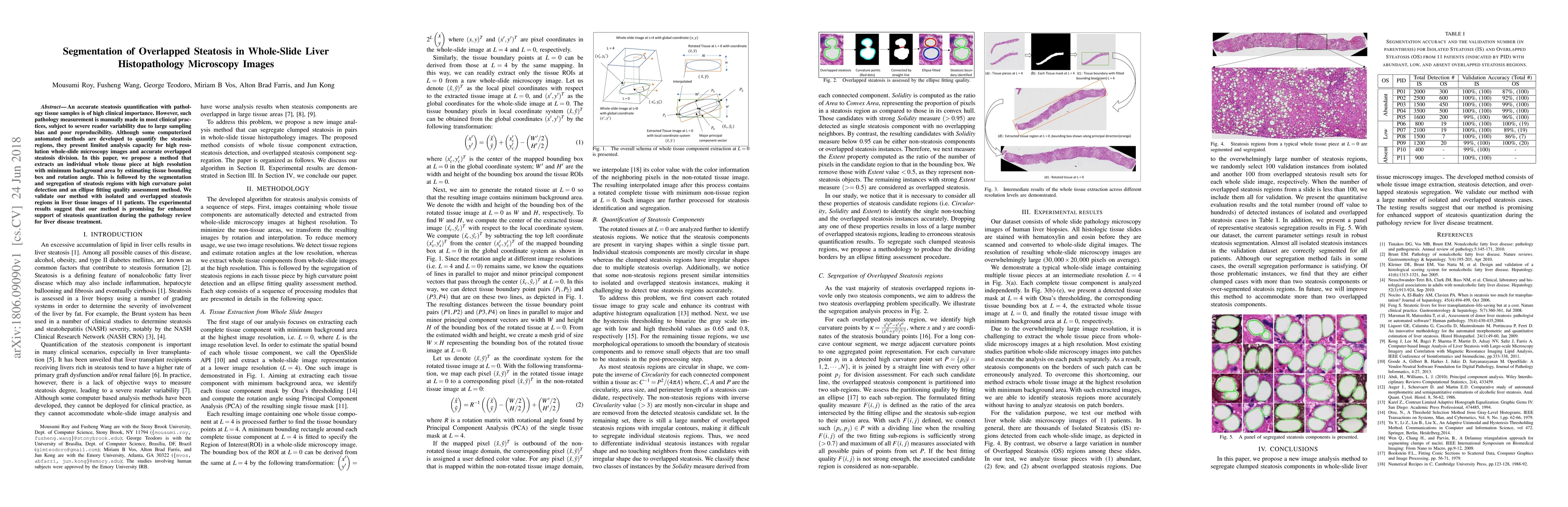
An accurate steatosis quantification with pathology tissue samples is of high clinical importance. However, such pathology measurement is manually made in most clinical practices, subject to severe reader variability due to large sampling bias and poor reproducibility. Although some computerized automated methods are developed to quantify the steatosis regions, they present limited analysis capacity for high resolution whole-slide microscopy images and accurate overlapped steatosis division. In this paper, we propose a method that extracts an individual whole tissue piece at high resolution with minimum background area by estimating tissue bounding box and rotation angle. This is followed by the segmentation and segregation of steatosis regions with high curvature point detection and an ellipse fitting quality assessment method. We validate our method with isolated and overlapped steatosis regions in liver tissue images of 11 patients. The experimental results suggest that our method is promising for enhanced support of steatosis quantization during the pathology review for liver disease treatment.
This graph shows how this paper is connected to other research. The current paper is shown in dark gray, papers that cite it are in green, and papers it references are in blue. You can:
• Hover over any node to see the paper title
• Click a node to view that paper's details
• Drag nodes to rearrange the graph
• Use the zoom controls (bottom right) to zoom in/out
Title | Authors | Year | Actions |
---|---|---|---|
An evolutionary algorithm-based optimization method for the classification and quantification of steatosis prevalence in liver biopsy images
|
Alexandros Bantaloukas-Arjmand, Vasileios Christou, Ioannis G. Tsoulos, M. Tsipouras, A. Tzallas, Ch Gogos, E. Glavas, N. Giannakeas
|
2021
|
View Paper |
Quantitative assessment of liver fibrosis by digital image analysis reveals correlation with qualitative clinical fibrosis staging in liver transplant patients
|
K. Jiang, M. Mohammad, W. Dar, Jun Kong, A. Farris
|
2020
|
View Paper |
Transfer Learning versus Custom CNN Architectures in NAFLD Biopsy Images
|
Alexandros Bantaloukas-Arjmand, Vasileios Christou, A. Tzallas, M. Tsipouras, C. T. Angelis, Georgios Tsoumanis, E. Glavas, R. Forlano, P. Manousou, N. Giannakeas
|
2020
|
View Paper |
Deep Learning Based Accurate Hepatic Steatosis Quantification for Histological Assessment of Liver Biopsies
|
M. Roy, Fusheng Wang, Hoang Vo, Dejun Teng, George Teodoro, A. Farris, Eduardo Castillo‐Leon, M. Vos, Jun Kong
|
2020
|
View Paper |
Whole Slide Imaging and Its Applications to Histopathological Studies of Liver Disorders
|
R. Melo, Maximilian W. D. Raas, Cinthia Palazzi, Vitor H Neves, Kássia K Malta, T. P. Silva
|
2020
|
View Paper |
Training of Deep Convolutional Neural Networks to Identify Critical Liver Alterations in Histopathology Image Samples
|
Alexandros Bantaloukas-Arjmand, C. T. Angelis, Vasileios Christou, A. Tzallas, M. Tsipouras, E. Glavas, R. Forlano, P. Manousou, N. Giannakeas
|
2019
|
View Paper |
Liver Steatosis Segmentation With Deep Learning Methods
|
Xiaoyuan Guo, Fusheng Wang, George Teodoro, A. Farris, Jun Kong
|
2019
|
View Paper |
Published: 2020-06-23
Amit Sethi, Garima Jain, Abhijeet Patil, Tripti Bameta, Swapnil Rane, Harsh Diwakar, Jay Sawant
Published: 2024-10-04
Comments (0)