Publication
Metrics
AI Quick Summary
This paper proposes a starlet wavelet-based algorithm for segmenting scanning electron microscopy images to locate gold nanoparticles in natural rubber membranes, achieving over 85% accuracy. The method aims to estimate nanoparticle density distribution and predict reduction kinetics in future studies.
Paper Preview
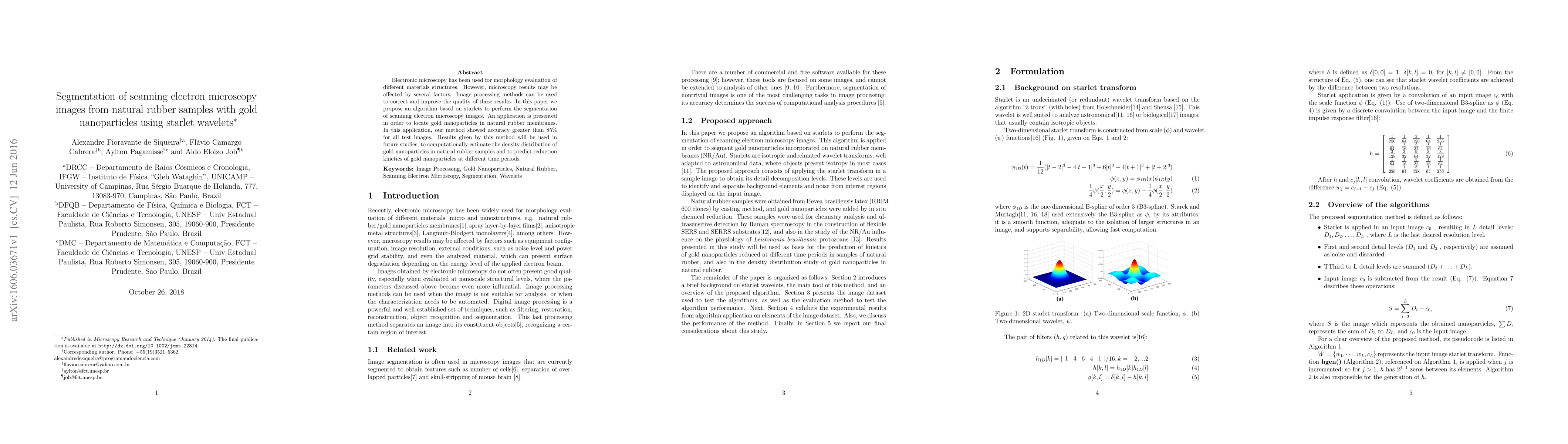
Abstract
Electronic microscopy has been used for morphology evaluation of different materials structures. However, microscopy results may be affected by several factors. Image processing methods can be used to correct and improve the quality of these results. In this paper we propose an algorithm based on starlets to perform the segmentation of scanning electron microscopy images. An application is presented in order to locate gold nanoparticles in natural rubber membranes. In this application, our method showed accuracy greater than 85% for all test images. Results given by this method will be used in future studies, to computationally estimate the density distribution of gold nanoparticles in natural rubber samples and to predict reduction kinetics of gold nanoparticles at different time periods.
AI Key Findings
Get AI-generated insights about this paper's methodology, results, significance, and more — seven facets brought into focus.
Impact
Paper Details
PDF Preview
Key Terms
Citation Network
Current paper (gray), citations (green), references (blue)
Display is limited for performance on very large graphs.
Discussion 0