Segmentation of Skeletal Muscle in Thigh Dixon MRI Based on Texture Analysis
Publication
Metrics
AI Quick Summary
This paper presents an automated segmentation method for skeletal muscle in thigh MRI using AdaBoost classification of texture features, including HOG and wavelet-based features, to address the limitations of manual segmentation. An atlas-based approach is also introduced, leveraging radiologist-provided ground truth for improved segmentation accuracy.
Paper Preview
Abstract
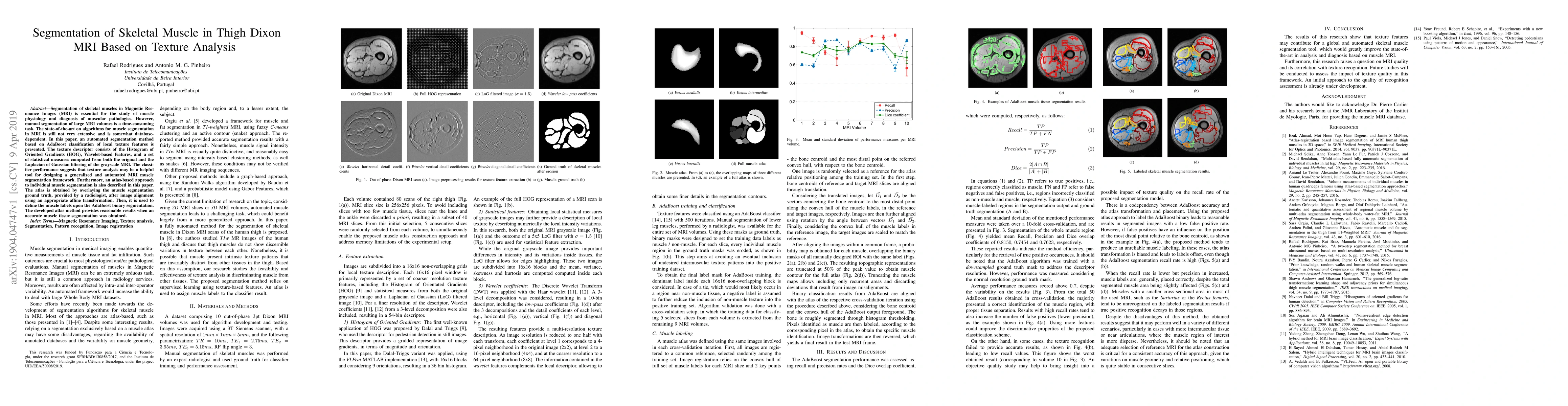
Segmentation of skeletal muscles in Magnetic Resonance Images (MRI) is essential for the study of muscle physiology and diagnosis of muscular pathologies. However, manual segmentation of large MRI volumes is a time-consuming task. The state-of-the-art on algorithms for muscle segmentation in MRI is still not very extensive and is somewhat database-dependent. In this paper, an automated segmentation method based on AdaBoost classification of local texture features is presented. The texture descriptor consists of the Histogram of Oriented Gradients (HOG), Wavelet-based features, and a set of statistical measures computed from both the original and the Laplacian of Gaussian filtering of the grayscale MRI. The classifier performance suggests that texture analysis may be a helpful tool for designing a generalized and automated MRI muscle segmentation framework. Furthermore, an atlas-based approach to individual muscle segmentation is also described in this paper. The atlas is obtained by overlaying the muscle segmentation ground truth, provided by a radiologist, after image alignment using an appropriate affine transformation. Then, it is used to define the muscle labels upon the AdaBoost binary segmentation. The developed atlas method provides reasonable results when an accurate muscle tissue segmentation was obtained.
AI Key Findings
Get AI-generated insights about this paper's methodology, results, significance, and more — seven facets brought into focus.
Impact
Paper Details
PDF Preview
Key Terms
Citation Network
Current paper (gray), citations (green), references (blue)
Display is limited for performance on very large graphs.
Discussion 0