Segmenting Cardiac Muscle Z-disks with Deep Neural Networks
Publication
Metrics
AI Quick Summary
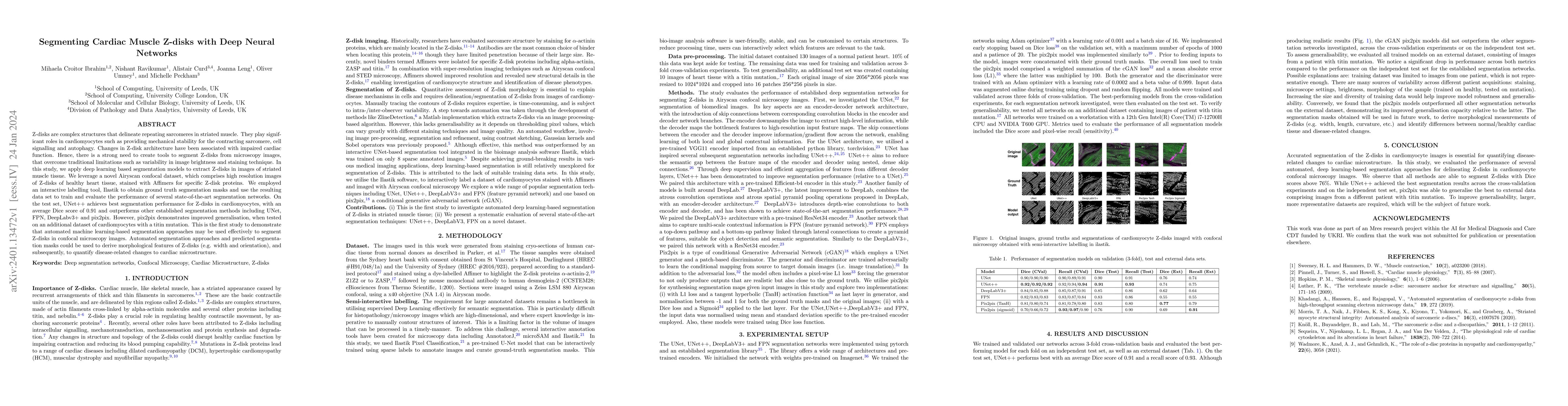
This study applies deep learning segmentation models to accurately delineate Z-disks in cardiac muscle tissue images, using a high-resolution Airyscan confocal dataset. UNet++ achieves the best performance with a Dice score of 0.91, while pix2pix shows improved generalization across different datasets.
Paper Preview
Abstract
Z-disks are complex structures that delineate repeating sarcomeres in striated muscle. They play significant roles in cardiomyocytes such as providing mechanical stability for the contracting sarcomere, cell signalling and autophagy. Changes in Z-disk architecture have been associated with impaired cardiac function. Hence, there is a strong need to create tools to segment Z-disks from microscopy images, that overcome traditional limitations such as variability in image brightness and staining technique. In this study, we apply deep learning based segmentation models to extract Z-disks in images of striated muscle tissue. We leverage a novel Airyscan confocal dataset, which comprises high resolution images of Z-disks of healthy heart tissue, stained with Affimers for specific Z-disk proteins. We employed an interactive labelling tool, Ilastik to obtain ground truth segmentation masks and use the resulting data set to train and evaluate the performance of several state-of-the-art segmentation networks. On the test set, UNet++ achieves best segmentation performance for Z-disks in cardiomyocytes, with an average Dice score of 0.91 and outperforms other established segmentation methods including UNet, FPN, DeepLabv3+ and pix2pix. However, pix2pix demonstrates improved generalisation, when tested on an additional dataset of cardiomyocytes with a titin mutation. This is the first study to demonstrate that automated machine learning-based segmentation approaches may be used effectively to segment Z-disks in confocal microscopy images. Automated segmentation approaches and predicted segmentation masks could be used to derive morphological features of Z-disks (e.g. width and orientation), and subsequently, to quantify disease-related changes to cardiac microstructure.
AI Key Findings
Get AI-generated insights about this paper's methodology, results, significance, and more — seven facets brought into focus.
Impact
Paper Details
Authors
PDF Preview
Key Terms
Citation Network
Current paper (gray), citations (green), references (blue)
Display is limited for performance on very large graphs.
Discussion 0