This paper proposes a deep learning method for accurately segmenting microcalcifications in mammograms, aiming to reduce false positives and improve detection accuracy. The approach focuses on training with hard pixels to enhance segmentation precision, facilitating better diagnosis and screening for Ductal Carcinoma in Situ.
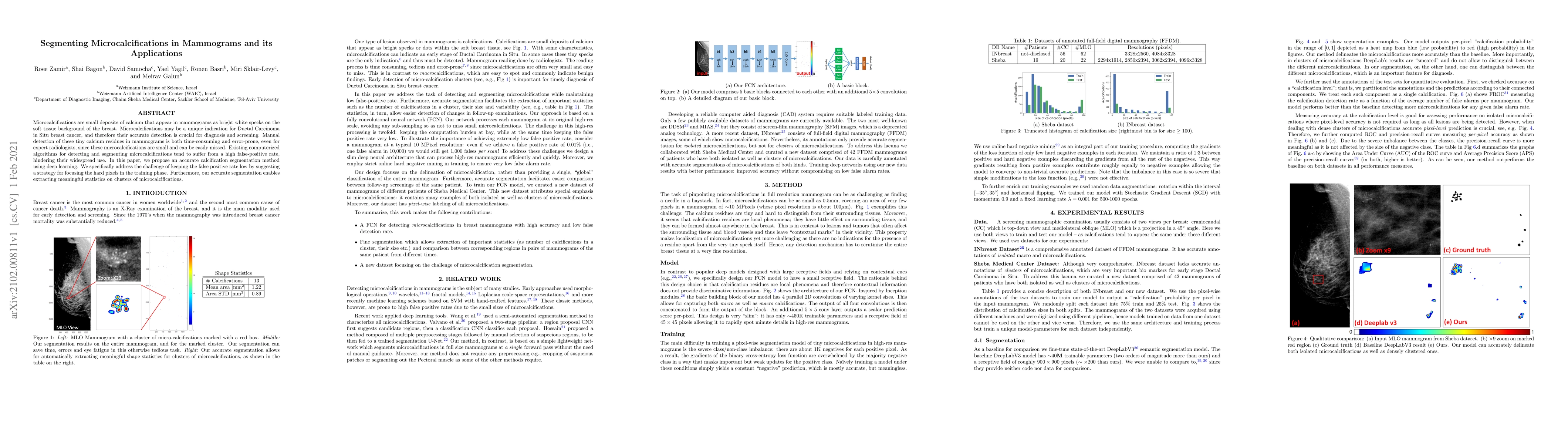
Microcalcifications are small deposits of calcium that appear in mammograms as bright white specks on the soft tissue background of the breast. Microcalcifications may be a unique indication for Ductal Carcinoma in Situ breast cancer, and therefore their accurate detection is crucial for diagnosis and screening. Manual detection of these tiny calcium residues in mammograms is both time-consuming and error-prone, even for expert radiologists, since these microcalcifications are small and can be easily missed. Existing computerized algorithms for detecting and segmenting microcalcifications tend to suffer from a high false-positive rate, hindering their widespread use. In this paper, we propose an accurate calcification segmentation method using deep learning. We specifically address the challenge of keeping the false positive rate low by suggesting a strategy for focusing the hard pixels in the training phase. Furthermore, our accurate segmentation enables extracting meaningful statistics on clusters of microcalcifications.
This graph shows how this paper is connected to other research. The current paper is shown in dark gray, papers that cite it are in green, and papers it references are in blue. You can:
• Hover over any node to see the paper title
• Click a node to view that paper's details
• Drag nodes to rearrange the graph
• Use the zoom controls (bottom right) to zoom in/out
Title | Authors | Year | Actions |
---|---|---|---|
A novel approach for segmentation and quantitative analysis of breast calcification in mammograms
|
Yunfei Tong, Jianrong Jiang, Fang Chen, Guanghua Guo, Chaoren Zhang, Tiana Deng
|
2024
|
View Paper |
Breast cancer diagnosis: A systematic review
|
Xin Wen, Xing Guo, Shuihua Wang, Zhihai Lu, Yu-dong Zhang
|
2024
|
View Paper |
Real-Time Pose Tracking for a Continuum Guidewire Robot Under Fluoroscopic Imaging
|
Sharan R. Ravigopal, Achraj Sarma, T. Brumfiel, J. Desai
|
2023
|
View Paper |
Towards Closed-loop Control of the Modified COAST Guidewire under Fluoroscopic Imaging for Endotracheal and Endovascular Interventions
|
Sharan R. Ravigopal, Kirsten M. Williams, J. Desai
|
2023
|
View Paper |
Real-time deep neural network-based automatic bowel gas segmentation on X-ray images for particle beam treatment
|
Toshio Kumakiri, S. Mori, Yasukuni Mori, R. Hirai, Ayato Hashimoto, Y. Tachibana, Hiroki Suyari, Hitoshi Ishikawa
|
2023
|
View Paper |
DeepMiCa: Automatic segmentation and classification of breast MIcroCAlcifications from mammograms
|
Alessia Gerbasi, Greta Clementi, F. Corsi, S. Albasini, A. Malovini, S. Quaglini, R. Bellazzi
|
2023
|
View Paper |
A survey of approaches in Deep Learning techniques for the detection and classification of mammography abnormalities
|
C. Flores, J. Ortega, Luis Antonio Salazar-Licea, M. Aceves-Fernández, Marzela Sánchez Osti
|
2022
|
View Paper |
Evaluation of different convolutional neural network encoder-decoder architectures for breast mass segmentation
|
A. Isosalo, H. Mustonen, Topi Turunen, P. Ipatti, J. Reponen, M. Nieminen, S. Inkinen
|
2022
|
View Paper |
Published: 2012-01-31
Ann-Katherine Carton, Clément Jailin, Serge Muller, Raoul de Sousa Silva, Ruben Sanchez de la Rosa
Published: 2022-11-09
Comments (0)