Self-DenseMobileNet employs advanced image techniques and a stacking-based meta-classifier to achieve high accuracy in lung nodule classification from chest radiographs, demonstrating robust performance internally and strong generalizability externally. The framework's interpretability is enhanced by class activation mapping.
No key terms available for this paper.
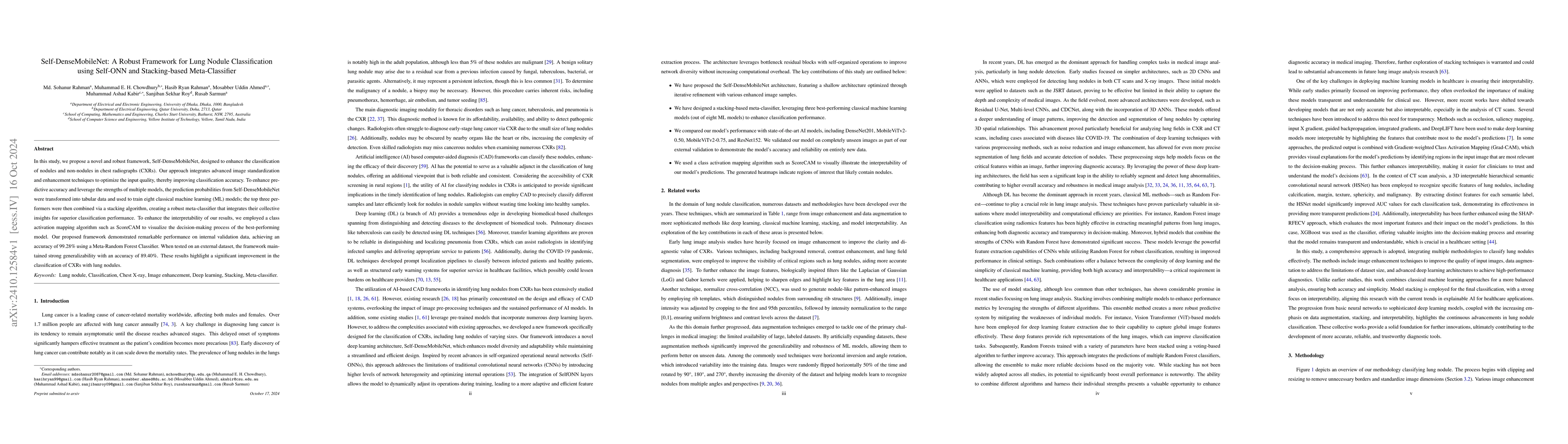
In this study, we propose a novel and robust framework, Self-DenseMobileNet, designed to enhance the classification of nodules and non-nodules in chest radiographs (CXRs). Our approach integrates advanced image standardization and enhancement techniques to optimize the input quality, thereby improving classification accuracy. To enhance predictive accuracy and leverage the strengths of multiple models, the prediction probabilities from Self-DenseMobileNet were transformed into tabular data and used to train eight classical machine learning (ML) models; the top three performers were then combined via a stacking algorithm, creating a robust meta-classifier that integrates their collective insights for superior classification performance. To enhance the interpretability of our results, we employed class activation mapping (CAM) to visualize the decision-making process of the best-performing model. Our proposed framework demonstrated remarkable performance on internal validation data, achieving an accuracy of 99.28\% using a Meta-Random Forest Classifier. When tested on an external dataset, the framework maintained strong generalizability with an accuracy of 89.40\%. These results highlight a significant improvement in the classification of CXRs with lung nodules.
This graph shows how this paper is connected to other research. The current paper is shown in dark gray, papers that cite it are in green, and papers it references are in blue. You can:
• Hover over any node to see the paper title
• Click a node to view that paper's details
• Drag nodes to rearrange the graph
• Use the zoom controls (bottom right) to zoom in/out
No citations found for this paper.
Title | Authors | Year | Actions |
---|---|---|---|
Nodule Detection and Generation on Chest X-Rays: NODE21 Challenge
|
Ecem Sogancioglu, B. Ginneken, Finn Behrendt, M. Bengs, Alexander Schlaefer, Miron Radu, Di Xu, Ke Sheng, Fabien Scalzo, Eric Marcus, Samuele Papa, Jonas Teuwen, E. Scholten, S. Schalekamp, N. Hendrix, C. Jacobs, Ward Hendrix, Clara I. S'anchez, K. Murphy
|
2024
|
View Paper |
Signer-Independent Arabic Sign Language Recognition System Using Deep Learning Model
|
K. Podder, Maymouna Ezeddin, M. Chowdhury, Md. Shaheenur Islam Sumon, A. Tahir, M. Ayari, Proma Dutta, A. Khandakar, Z. Mahbub, M. A. Kadir
|
2023
|
View Paper |
Medical image data augmentation: techniques, comparisons and interpretations
|
Evgin Göçeri
|
2023
|
View Paper |
Computer-Aided Diagnosis of COVID-19 from Chest X-ray Images Using Hybrid-Features and Random Forest Classifier
|
Kashif Shaheed, P. Szczuko, Qaisar Abbas, Ayyaz Hussain, Mubarak Albathan
|
2023
|
View Paper |
Self-attention random forest for breast cancer image classification
|
Jia Li, Jingwen Shi, Jianrong Chen, Z. Du, Li Huang
|
2023
|
View Paper |
CADNet157 model: fine-tuned ResNet152 model for breast cancer diagnosis from mammography images
|
Raouia Mokni, Mariem Haoues
|
2022
|
View Paper |
CVNets: High Performance Library for Computer Vision
|
Sachin Mehta, Farzad Abdolhosseini, Mohammad Rastegari
|
2022
|
View Paper |
Visual attribution for deep learning segmentation in medical imaging
|
S. Mullan, M. Sonka
|
2022
|
View Paper |
Explainable Machine Learning for COVID-19 Pneumonia Classification With Texture-Based Features Extraction in Chest Radiography
|
L. Moura, C. M. Oliveira, C. Dartora, Rodrigo C. Barros, A. M. D. Silva
|
2022
|
View Paper |
A Study on the Combination of Image Preprocessing Method Based on Texture Feature and Segmentation Algorithm for Breast Ultrasound Images
|
Senxin Cai, Yifeng Zhu, Jing Zhang, Tong Liu
|
2022
|
View Paper |
MobileViT: Light-weight, General-purpose, and Mobile-friendly Vision Transformer
|
Sachin Mehta, Mohammad Rastegari
|
2021
|
View Paper |
Weakly supervised segmentation of tumor lesions in PET-CT hybrid imaging
|
Marcel Früh, Marc Fischer, A. Schilling, S. Gatidis, Tobias Hepp
|
2021
|
View Paper |
Automatic and Reliable Leaf Disease Detection Using Deep Learning Techniques
|
M. Chowdhury, Tawsifur Rahman, A. Khandakar, M. Ayari, Aftab Khan, Muhammad Salman Khan, N. Al-Emadi, M. Reaz, M. Islam, S. Ali
|
2021
|
View Paper |
COVID-19 infection localization and severity grading from chest X-ray images
|
A. Tahir, M. Chowdhury, A. Khandakar, Tawsifur Rahman, Yazan Qiblawey, U. Khurshid, S. Kiranyaz, Nabil Ibtehaz, M. S. Rahman, Somaya Al-Madeed, K. Hameed, T. Hamid, S. Mahmud, Maymouna Ezeddin
|
2021
|
View Paper |
Automatic Classification and Accuracy by Deep Learning Using CNN Methods in Lung Chest X-Ray Images
|
V. Thamilarasi, R. Roselin
|
2021
|
View Paper |
VinDr-CXR: An open dataset of chest X-rays with radiologist’s annotations
|
H. Nguyen, K. Lam, Linh T. Le, Hieu Pham, Dat Q. Tran, D. Nguyen, Dung D. Le, Chi M. Pham, Han Tong, D. H. Dinh, C. D. Do, L. T. Doan, Cuong N. Nguyen, Binh T. Nguyen, Que V. Nguyen, A. D. Hoang, H. N. Phan, A. Nguyen, P. Ho, D. Ngo, N. T. Nguyen, N. T. Nguyen, Minh-Son Dao, V. Vu
|
2020
|
View Paper |
An Image is Worth 16x16 Words: Transformers for Image Recognition at Scale
|
Alexey Dosovitskiy, Lucas Beyer, Alexander Kolesnikov, Dirk Weissenborn, Xiaohua Zhai, Thomas Unterthiner, Mostafa Dehghani, Matthias Minderer, G. Heigold, S. Gelly, Jakob Uszkoreit, N. Houlsby
|
2020
|
View Paper |
COVID-19 infection map generation and detection from chest X-ray images
|
A. Degerli, Mete Ahishali, Mehmet Yamaç, S. Kiranyaz, M. Chowdhury, Khalid Hameed, T. Hamid, R. Mazhar, M. Gabbouj
|
2020
|
View Paper |
Self-Organized Operational Neural Networks for Severe Image Restoration Problems
|
Junaid Malik, S. Kiranyaz, M. Gabbouj
|
2020
|
View Paper |
Contrast Enhancement of Low-Contrast Medical Images Using Modified Contrast Limited Adaptive Histogram Equalization
|
S. Khan, Shariq Hussain, Shunkun Yang
|
2020
|
View Paper |
A Novel Data Augmentation Method Using Style-Based GAN for Robust Pulmonary Nodule Segmentation
|
Haoqi Shi, Junguo Lu, Qianjun Zhou
|
2020
|
View Paper |
A robust convolutional neural network for lung nodule detection in the presence of foreign bodies
|
M. Schultheiss, Sebastian A. Schober, Marie Lodde, J. Bodden, Juliane Aichele, C. Müller-Leisse, B. Renger, F. Pfeiffer, D. Pfeiffer
|
2020
|
View Paper |
Reliable Tuberculosis Detection Using Chest X-Ray With Deep Learning, Segmentation and Visualization
|
Tawsifur Rahman, A. Khandakar, M. A. Kadir, K. R. Islam, Khandaker F. Islam, R. Mazhar, T. Hamid, M. Islam, Z. Mahbub, M. Ayari, M. Chowdhury
|
2020
|
View Paper |
A combination of FractalNet and CNN for Lung Nodule Classification
|
Amrita Naik, D. Edla, Venkatanareshbabu Kuppili
|
2020
|
View Paper |
Exploration of Interpretability Techniques for Deep COVID-19 Classification Using Chest X-ray Images
|
S. Chatterjee, Fatima Saad, Chompunuch Sarasaen, Suhita Ghosh, Rupali Khatun, P. Radeva, G. Rose, Sebastian Stober, O. Speck, A. Nürnberger
|
2020
|
View Paper |
Pre‐eclampsia‐like syndrome induced by severe COVID‐19: a prospective observational study
|
M. Mendoza, I. García-Ruiz, N. Maíz, C. Rodó, P. Garcia-Manau, B. Serrano, R. Lopez-Martinez, J. Balcells, N. Fernández-hidalgo, E. Carreras, A. Suy
|
2020
|
View Paper |
Pulmonary nodule detection on chest radiographs using balanced convolutional neural network and classic candidate detection
|
Sheng Chen, Yaqi Han, Jinqiu Lin, Xiangyu Zhao, Ping Kong
|
2020
|
View Paper |
Data Preprocessing Via Compositions Multi-Channel MRI Images to Improve Brain Tumor Segmentation
|
N. Tolstokulakov, Evgeny Nikolaevich Pavlovskiy, B. Tuchinov, E. Amelina, M. Amelin, A. Letyagin, S. Golushko, V. Groza
|
2020
|
View Paper |
Thorax-Net: An Attention Regularized Deep Neural Network for Classification of Thoracic Diseases on Chest Radiography
|
Hongyu Wang, Haozhe Jia, Le Lu, Yong Xia
|
2020
|
View Paper |
Linear Discriminant Analysis
|
Hong Zhou
|
2020
|
View Paper |
Score-CAM: Score-Weighted Visual Explanations for Convolutional Neural Networks
|
Haofan Wang, Zifan Wang, Mengnan Du, Fan Yang, Zijian Zhang, Sirui Ding, Piotr (Peter) Mardziel, Xia Hu
|
2019
|
View Paper |
A survey on Image Data Augmentation for Deep Learning
|
Connor Shorten, T. Khoshgoftaar
|
2019
|
View Paper |
Deep Feature Learning from a Hospital-Scale Chest X-ray Dataset with Application to TB Detection on a Small-Scale Dataset
|
Ophir Gozes, H. Greenspan
|
2019
|
View Paper |
Operational neural networks
|
S. Kiranyaz, T. Ince, Alexandros Iosifidis, M. Gabbouj
|
2019
|
View Paper |
PadChest: A large chest x-ray image dataset with multi-label annotated reports
|
A. Bustos, A. Pertusa, J. M. Salinas, M. Iglesia-Vayá
|
2019
|
View Paper |
Diagnosis and management of peripheral lung nodule.
|
Taha Khan, Y. Usman, T. Abdo, F. Chaudry, J. Keddissi, H. Youness
|
2019
|
View Paper |
Multi-Scale Dense Networks for Deep High Dynamic Range Imaging
|
Qingsen Yan, Dong Gong, Pingping Zhang, Javen Qinfeng Shi, Jinqiu Sun, I. Reid, Yanning Zhang
|
2019
|
View Paper |
Global cancer statistics 2018: GLOBOCAN estimates of incidence and mortality worldwide for 36 cancers in 185 countries
|
F. Bray, J. Ferlay, I. Soerjomataram, R. Siegel, Lindsey A. Torre, A. Jemal
|
2018
|
View Paper |
Automatic Lung Cancer Prediction from Chest X-ray Images Using the Deep Learning Approach
|
Worawate Ausawalaithong, S. Marukatat, Arjaree Thirach, Theerawit Wilaiprasitporn
|
2018
|
View Paper |
Using Multi-level Convolutional Neural Network for Classification of Lung Nodules on CT images
|
Juan Lyu, S. Ling
|
2018
|
View Paper |
RUN: Residual U-Net for Computer-Aided Detection of Pulmonary Nodules without Candidate Selection
|
Tian Lan, Yuanyuan Li, Jonah Kimani Murugi, Yi Ding, Zhiguang Qin
|
2018
|
View Paper |
Identifying Medical Diagnoses and Treatable Diseases by Image-Based Deep Learning
|
Daniel S. Kermany, Daniel S. Kermany, M. Goldbaum, Wenjia Cai, C. Valentim, Huiying Liang, Sally L. Baxter, A. McKeown, Ge Yang, Xiaokang Wu, Fangbing Yan, Justin Dong, Made K. Prasadha, Jacqueline Pei, Jacqueline Pei, M. Y. Ting, Jie Zhu, Christina M. Li, Sierra Hewett, Sierra Hewett, Jason Dong, Ian Ziyar, Alexander Shi, Runze Zhang, Lianghong Zheng, Rui Hou, W. Shi, Xin Fu, Xin Fu, Yaou Duan, V. N. Huu, V. N. Huu, Cindy Wen, Edward Zhang, Edward Zhang, Charlotte L. Zhang, Charlotte L. Zhang, Oulan Li, Oulan Li, Xiaobo Wang, M. Singer, Xiaodong Sun, Jie Xu, A. Tafreshi, M. Lewis, H. Xia, Kang Zhang
|
2018
|
View Paper |
Tuberculosis Detection from Chest Radiographs: A Comprehensive Survey on Computer-Aided Diagnosis Techniques
|
Rahul Hooda, Ajay Mittal, S. Sofat
|
2017
|
View Paper |
A Novel Image Classification Method with CNN-XGBoost Model
|
Xudie Ren, Haonan Guo, Shenghong Li, Shilin Wang, Jianhua Li
|
2017
|
View Paper |
SmoothGrad: removing noise by adding noise
|
D. Smilkov, Nikhil Thorat, Been Kim, F. Viégas, M. Wattenberg
|
2017
|
View Paper |
[Pulmonary nodules].
|
R. Prevel, M. Receveur, A. Duvignaud
|
2017
|
View Paper |
Grad-CAM: Visual Explanations from Deep Networks via Gradient-Based Localization
|
Ramprasaath R. Selvaraju, Abhishek Das, Ramakrishna Vedantam, Michael Cogswell, Devi Parikh, Dhruv Batra
|
2016
|
View Paper |
Using grayscale images for object recognition with convolutional-recursive neural network
|
H. Bui, M. Lech, E. Cheng, K. Neville, I. Burnett
|
2016
|
View Paper |
Epidemiology of Lung Cancer.
|
Y. Mao, Ding Yang, Jie He, M. Krasna
|
2016
|
View Paper |
Deep Residual Learning for Image Recognition
|
Kaiming He, X. Zhang, Shaoqing Ren, Jian Sun
|
2015
|
View Paper |
Localized Energy-Based Normalization of Medical Images: Application to Chest Radiography
|
R. Philipsen, P. Maduskar, L. Hogeweg, Jaime Melendez, C. I. Sánchez, B. Ginneken
|
2015
|
View Paper |
Histogram Equalization Techniques for Contrast Enhancement: A Review
|
Sourav Das, T. Gulati, Vikas Mittal
|
2015
|
View Paper |
A robust SVM classification framework using PSM for multi-class recognition
|
Jinhui Chen, T. Takiguchi, Y. Ariki
|
2015
|
View Paper |
Review of Various Histogram Based Medical Image Enhancement Techniques
|
H. N. Vidyasaraswathi, M. C. Hanumantharaju
|
2015
|
View Paper |
Computer-aided detection improves detection of pulmonary nodules in chest radiographs beyond the support by bone-suppressed images.
|
S. Schalekamp, B. van Ginneken, Emmeline Koedam, M. Snoeren, A. Tiehuis, R. Wittenberg, N. Karssemeijer, C. Schaefer-Prokop
|
2014
|
View Paper |
Gradient boosting machines, a tutorial
|
Alexey Natekin, A. Knoll
|
2013
|
View Paper |
Design and Development of a Multimodal Biomedical Information Retrieval System
|
Dina Demner-Fushman, Sameer Kiran Antani, Matthew S. Simpson, G. Thoma
|
2012
|
View Paper |
Diagnosis of Pneumothorax by Radiography and Ultrasonography: A Meta-analysis
|
C. Johnston
|
2012
|
View Paper |
A Survey on Transfer Learning
|
Sinno Jialin Pan, Qiang Yang
|
2010
|
View Paper |
Pulmonary nodules: detection, assessment, and CAD.
|
F. Girvin, J. Ko
|
2008
|
View Paper |
Neural networks for computer-aided diagnosis: detection of lung nodules in chest radiograms
|
G. Coppini, S. Diciotti, M. Falchini, N. Villari, G. Valli
|
2003
|
View Paper |
Handbook of Medical Imaging, Processing and Analysis
|
A. Fenster
|
2001
|
View Paper |
Support vector machine learning algorithm and transduction
|
A. Gamermann
|
2000
|
View Paper |
A research insight toward the significance in extraction of retinal blood vessels from fundus images and its various implementations
|
Nimisha Anns Oommen, P. Darsana
|
2022
|
View Paper |
Labeling Chest X-Ray Reports Using Deep Learning
|
Maram Mahmoud A. Monshi, Josiah Poon, Yuk Ying Chung, F. M. Monshi
|
2021
|
View Paper |
Medical Image Classification Using a Light-Weighted Hybrid Neural Network Based on PCANet and DenseNet
|
Zhiwen Huang, Xingxing Zhu, Mingyue Ding, Xuming Zhang
|
2020
|
View Paper |
Medical image enhancement based on histogram algorithms
|
Nema Salem, Hebatullah M. Malik, Asmaa Shams
|
2019
|
View Paper |
Multilayer Perceptron (MLP)
|
H. Taud, J. Mas
|
2018
|
View Paper |
An objective method to identify optimum clip-limit and histogram specification of contrast limited adaptive histogram equalization for MR images
|
Justin Joseph, J. Sivaraman, R. Periyasamy, V. R. Simi
|
2017
|
View Paper |
ChestX-Ray8: Hospital-Scale
|
2017
|
||
Lung Nodule Detection in X-Ray Images: A New Feature Set
|
B. B. Oğul, P. Koşucu, Ahmet Özçam, S. D. Kanik
|
2015
|
View Paper |
Explaining AdaBoost
|
R. Schapire
|
2013
|
View Paper |
Development of a digital image database for chest radiographs with and without a lung nodule: receiver operating characteristic analysis of radiologists' detection of pulmonary nodules.
|
J. Shiraishi, S. Katsuragawa, J. Ikezoe, Tsuneo Matsumoto, Takeshi Kobayashi, K. Komatsu, Mitate Matsui, H. Fujita, Y. Kodera, K. Doi
|
2000
|
View Paper |
Missed lung cancer on chest radiography and computed tomography: imaging and medicolegal issues.
|
Charles S. White, Ari I. Salis, Cristopher A Meyer
|
1999
|
View Paper |
Artificial convolution neural network techniques and applications for lung nodule detection
|
S. Lo, S. Lou, Jyh-Shyan Lin, M. Freedman, M. Chien, S. Mun
|
1995
|
View Paper |
2023. An Interpretable Three-Dimensional Artificial Intelligence Model for Computer-Aided Diagnosis of Lung Nodules in Computed Tomography Images
|
|||
2023. Cdc net: multi-classification convolutional neural network model for detection of covid-19, pneumothorax, pneumonia, lung cancer, and tuberculosis using chest x-rays
|
|||
Rethinking the transfer learning architecture for respiratory diseases and covid-19 diagnosis
|
Showing first page preview. Click "Open Full PDF" to view the complete paper.
Muhammad Usman, Sung Hyun Kim, Siddique Latif, Azka Rehman, Abdullah Shahid, Tariq Mahmood Khan, Shi Sub Byon, Yeong Gil Shin
Published: 2023-04-05
Yulei Qin, Yun Gu, Liang Chen, Minghui Zhang, Feng Yao, Hanxiao Zhang, Guang-Zhong Yang, Xiao Gu, Zhexin Wang
Published: 2022-02-28
Jiahao Lu, Sune Darkner, Kenny Erleben, Oswin Krause, Chong Yin, Michael Bachmann Nielsen
Published: 2022-11-08
You need to log in to post a comment.
Your comment contains inappropriate language.
Are you sure you want to remove this tag?
Someone just favorited a paper!
Comments (0)