Self-Supervised ImageNet Representations for In Vivo Confocal Microscopy: Tortuosity Grading without Segmentation Maps
Publication
Metrics
Paper Preview
Abstract
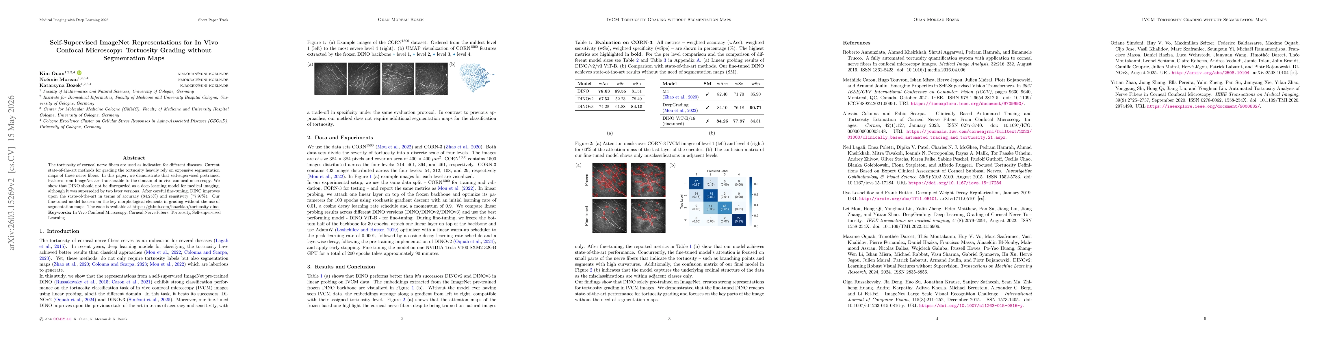
The tortuosity of corneal nerve fibers are used as indication for different diseases. Current state-of-the-art methods for grading the tortuosity heavily rely on expensive segmentation maps of these nerve fibers. In this paper, we demonstrate that self-supervised pretrained features from ImageNet are transferable to the domain of in vivo confocal microscopy. We show that DINO should not be disregarded as a deep learning model for medical imaging, although it was superseded by two later versions. After careful fine-tuning, DINO improves upon the state-of-the-art in terms of accuracy (84,25%) and sensitivity (77,97%). Our fine-tuned model focuses on the key morphological elements in grading without the use of segmentation maps.
AI Key Findings
Get AI-generated insights about this paper's methodology, results, significance, and more — seven facets brought into focus.
Discussion 0