This paper introduces a self-supervised method for nuclei segmentation in histopathological images, leveraging magnification level identification to generate a preliminary self-supervision signal. The approach outperforms other unsupervised methods and matches the performance of supervised ones on the MoNuSeg dataset.
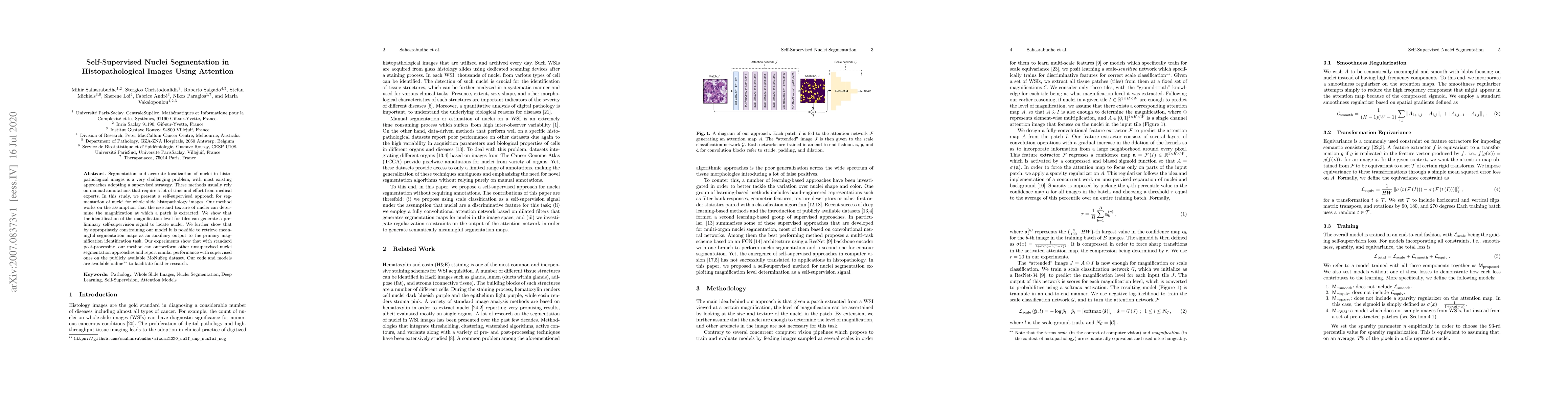
Segmentation and accurate localization of nuclei in histopathological images is a very challenging problem, with most existing approaches adopting a supervised strategy. These methods usually rely on manual annotations that require a lot of time and effort from medical experts. In this study, we present a self-supervised approach for segmentation of nuclei for whole slide histopathology images. Our method works on the assumption that the size and texture of nuclei can determine the magnification at which a patch is extracted. We show that the identification of the magnification level for tiles can generate a preliminary self-supervision signal to locate nuclei. We further show that by appropriately constraining our model it is possible to retrieve meaningful segmentation maps as an auxiliary output to the primary magnification identification task. Our experiments show that with standard post-processing, our method can outperform other unsupervised nuclei segmentation approaches and report similar performance with supervised ones on the publicly available MoNuSeg dataset. Our code and models are available online to facilitate further research.
Get AI-generated insights about this paper's methodology, results, and significance.
Current paper (gray), citations (green), references (blue)
Mausam, Aayush Kumar Tyagi, Vaibhav Mishra et al.
Ayush Roy, Ram Sarkar, Payel Pramanik et al.
Prathosh A. P, Seshan Srirangarajan, Suhas Srinath et al.
Lei Fan, Yang Song, Maurice Pagnucco et al.
Title | Authors | Year | Actions |
---|---|---|---|
HistoMSC: Density and topology analysis for AI-based visual annotation of histopathology whole slide images
|
Zahoor Ahmad, Khaled A. Al-Thelaya, M. Alzubaidi, Faaiz Joad, N. Gilal, William Mifsud, Sabri Boughorbel, G. Pintore, Enrico Gobbetti, Jens Schneider, Marco Agus
|
2025
|
View Paper |
COIN: Confidence Score-Guided Distillation for Annotation-Free Cell Segmentation
|
Sang-Kee Jo, Seo Jin Lee, Seungwoo Lee, Seohyung Hong, Hyungseok Seo, Kyungsu Kim
|
2025
|
View Paper |
Prompt as Knowledge Bank: Boost Vision-language model via Structural Representation for zero-shot medical detection
|
Yuguang Yang, Tongfei Chen, Haoyu Huang, Linlin Yang, Chunyu Xie, Dawei Leng, Xianbin Cao, Baochang Zhang
|
2025
|
View Paper |
CompSegNet: An enhanced U-shaped architecture for nuclei segmentation in H&E histopathology images
|
Mohamed Traoré, Emrah Hançer, R. Samet, Z. Yildirim, Nooshin Nemati
|
2024
|
View Paper |
AttriPrompter: Auto-Prompting With Attribute Semantics for Zero-Shot Nuclei Detection via Visual-Language Pre-Trained Models
|
Yongjian Wu, Yang Zhou, Jiya Saiyin, Bingzheng Wei, Maode Lai, Jianzhong Shou, Yan Xu
|
2024
|
View Paper |
A Survey on Cell Nuclei Instance Segmentation and Classification: Leveraging Context and Attention
|
João D. Nunes, D. Montezuma, Domingos Oliveira, Tania Pereira, Jaime S. Cardoso
|
2024
|
View Paper |
A Survey on Cell Nuclei Instance Segmentation and Classification: Leveraging Context and Attention
|
João D. Nunes, D. Montezuma, Domingos Oliveira, Tania Pereira, Jaime S. Cardoso
|
2024
|
View Paper |
A-ReSEUnet: Achieve no-label binary segmentation of nuclei in histology images
|
Jui-Hung Chang, Pei-Hsuan Wu, Ti-Hao Wang, Pau-Choo Chung
|
2024
|
View Paper |
PathoDuet: Foundation models for pathological slide analysis of H&E and IHC stains
|
Shengyi Hua, Fang Yan, Tianle Shen, Lei Ma, Xiaofan Zhang
|
2024
|
View Paper |
Mapping the landscape of histomorphological cancer phenotypes using self-supervised learning on unannotated pathology slides
|
Adalberto Claudio Quiros, N. Coudray, A. Yeaton, Xinyu Yang, Bojing Liu, H. Le, Luis Chiriboga, A. Karimkhan, Navneet Narula, David A Moore, Christopher Y. Park, H. Pass, Andre L Moreira, J. Le Quesne, A. Tsirigos, Ke Yuan
|
2024
|
View Paper |
Diffusion Models for Nuclei Segmentation in Low Data Regimes
|
Konstantinos Alexis, S. Christodoulidis, Dimitrios Gunopulos, M. Vakalopoulou
|
2024
|
View Paper |
Cluster-based histopathology phenotype representation learning by self-supervised multi-class-token hierarchical ViT
|
Jiarong Ye, Shivam Kalra, Mohammad Saleh Miri
|
2024
|
View Paper |
Computational pathology: A survey review and the way forward
|
M. S. Hosseini, B. Bejnordi, Vincent Quoc-Huy Trinh, Lyndon Chan, Danial Hasan, Xingwen Li, Stephen Yang, Taehyo Kim, Haochen Zhang, Theodore Wu, Kajanan Chinniah, Sina Maghsoudlou, Ryan Zhang, Jiadai Zhu, Samir Khaki, Andrei Buin, Fatemeh Chaji, Ala Salehi, Bich Ngoc Nguyen, Dimitris Samaras, K. Plataniotis
|
2024
|
View Paper |
Enhancing breast cancer classification via histopathological image analysis: Leveraging self-supervised contrastive learning and transfer learning
|
Faisal Bin Ashraf, S.M. Maksudul Alam, Shahriar M. Sakib
|
2024
|
View Paper |
Attention De-sparsification Matters: Inducing Diversity in Digital Pathology Representation Learning
|
S. Kapse, Srijan Das, Jingwei Zhang, Rajarsi R. Gupta, J. Saltz, D. Samaras, P. Prasanna
|
2023
|
View Paper |
Breast Cancer Histopathological Images Segmentation Using Deep Learning
|
Wafaa Rajaa Drioua, Nacéra Benamrane, L. Sais
|
2023
|
View Paper |
A Comprehensive Overview of Computational Nuclei Segmentation Methods in Digital Pathology
|
Vasileios Magoulianitis, Catherine A. Alexander, C.-C. Jay Kuo
|
2023
|
View Paper |
Semi-Supervised Semantic Segmentation of Cell Nuclei via Diffusion-based Large-Scale Pre-Training and Collaborative Learning
|
Zhucheng Shao, S. Sengupta, Hua Li, M. Anastasio
|
2023
|
View Paper |
Nested star-shaped objects segmentation using diameter annotations
|
Robin Camarasa, H. Kervadec, M. Kooi, J. Hendrikse, P. Nederkoorn, Daniel Bos, Marleen de Bruijne
|
2023
|
View Paper |
All-in-SAM: from Weak Annotation to Pixel-wise Nuclei Segmentation with Prompt-based Finetuning
|
C. Cui, Ruining Deng, Quan Liu, Tianyuan Yao, Shunxing Bao, Lucas W. Remedios, Yucheng Tang, Yuankai Huo
|
2023
|
View Paper |
Zero-shot Nuclei Detection via Visual-Language Pre-trained Models
|
Yongjian Wu, Yangqiaoyu Zhou, Jiya Saiyin, Bingzheng Wei, Maode Lai, Jianzhong Shou, Yubo Fan, Yan Xu
|
2023
|
View Paper |
Deep Learning in Breast Cancer Imaging: A Decade of Progress and Future Directions
|
Luyang Luo, Xi Wang, Yi-Mou Lin, Xiaoqi Ma, Andong Tan, R. Chan, V. Vardhanabhuti, W. C. Chu, Kwang-Ting Cheng, Hao Chen
|
2023
|
View Paper |
Label-Efficient Deep Learning in Medical Image Analysis: Challenges and Future Directions
|
Cheng Jin, Zhengrui Guo, Yi-Mou Lin, Luyang Luo, Hao Chen
|
2023
|
View Paper |
A Review of Predictive and Contrastive Self-supervised Learning for Medical Images
|
Wei-Chien Wang, E. Ahn, Da-wei Feng, Jinman Kim
|
2023
|
View Paper |
Artifact Removal in Histopathology Images
|
Cameron Dahan, S. Christodoulidis, M. Vakalopoulou, Joseph Boyd
|
2022
|
View Paper |
Which Pixel to Annotate: A Label-Efficient Nuclei Segmentation Framework
|
Wei Lou, Haofeng Li, Guanbin Li, Xiaoguang Han, Xiang Wan
|
2022
|
View Paper |
An Unsupervised Parameter-Free Nuclei Segmentation Method for Histology Images
|
Vasileios Magoulianitis, Peida Han, Yijing Yang, C. J. Kuo
|
2022
|
View Paper |
A Self-Supervised Learning Based Framework for Eyelid Malignant Melanoma Diagnosis in Whole Slide Images
|
Zijing Jiang, Linyan Wang, Yaqi Wang, Gangyong Jia, G. Zeng, Jun Wang, Yunxiang Li, Dechao Chen, Guiping Qian, Qun Jin
|
2022
|
View Paper |
Nuclei segmentation of HE stained histopathological images based on feature global delivery connection network
|
Peng Shi, Jing Zhong, Liyan Lin, Lin Lin, Huachang Li, Chongshu Wu
|
2022
|
View Paper |
HistoPerm: A permutation-based view generation approach for improving histopathologic feature representation learning
|
Joseph DiPalma, L. Torresani, S. Hassanpour
|
2022
|
View Paper |
Nuclei and glands instance segmentation in histology images: a narrative review
|
Esha Sadia Nasir, Arshi Parvaiz, M. Fraz
|
2022
|
View Paper |
Efficient Self-Supervision using Patch-based Contrastive Learning for Histopathology Image Segmentation
|
Nicklas Boserup, Raghavendra Selvan
|
2022
|
View Paper |
Towards label-efficient automatic diagnosis and analysis: a comprehensive survey of advanced deep learning-based weakly-supervised, semi-supervised and self-supervised techniques in histopathological image analysis
|
Linhao Qu, Siyu Liu, Xiaoyu Liu, Manning Wang, Zhijian Song
|
2022
|
View Paper |
A survey on artificial intelligence in histopathology image analysis
|
M. Abdelsamea, Usama Zidan, Zakaria Senousy, M. Gaber, E. Rakha, Mohammad Ilyas
|
2022
|
View Paper |
NormToRaw: A Style Transfer Based Self-supervised Learning Approach for Nuclei Segmentation
|
Xianlai Chen, Xuantong Zhong, Taixiang Li, Ying An, L. Mo
|
2022
|
View Paper |
CS-CO: A Hybrid Self-Supervised Visual Representation Learning Method for H&E-stained Histopathological Images
|
Pengshuai Yang, Xiaoxu Yin, Haiming Lu, Zhongliang Hu, Xuegong Zhang, Rui Jiang, Hairong Lv
|
2022
|
View Paper |
Transformer-based unsupervised contrastive learning for histopathological image classification
|
Xiyue Wang, Sen Yang, Jun Zhang, Minghui Wang, Jing Zhang, Wei Yang, Junzhou Huang, Xiao Han
|
2022
|
View Paper |
Mapping the landscape of histomorphological cancer phenotypes using self-supervised learning on unlabeled, unannotated pathology slides
|
A. Quiros, N. Coudray, A. Yeaton, Xinyu Yang, Bojing Liu, H. Le, L. Chiriboga, A. Karimkhan, N. Narula, David A Moore, Christopher Y. Park, H. Pass, A. Moreira, J. L. Quesne, A. Tsirigos, Ke-Fei Yuan
|
2022
|
View Paper |
Deep learning – promises for 3D nuclear imaging: a guide for biologists
|
Guillaume Mougeot, Tristan Dubos, F. Chausse, E. Péry, K. Graumann, C. Tatout, D. Evans, S. Desset
|
2022
|
View Paper |
HUNIS: High-Performance Unsupervised Nuclei Instance Segmentation
|
Vasileios Magoulianitis, Yijing Yang, C.-C. Jay Kuo
|
2022
|
View Paper |
MixCL: Pixel label matters to contrastive learning
|
Jun Li, Quan Quan, S. K. Zhou
|
2022
|
View Paper |
Scribble-Supervised Meibomian Glands Segmentation in Infrared Images
|
Xiaoming Liu, Shuo Wang, Ying Zhang, Quan Yuan
|
2022
|
View Paper |
Recent Advances of Deep Learning for Computational Histopathology: Principles and Applications
|
Yawen Wu, Michael Cheng, Shuo Huang, Zongxiang Pei, Yi Zuo, Jianxin Liu, Kai Yang, Qi Zhu, J. Zhang, Honghai Hong, Daoqiang Zhang, Kun Huang, Liang Cheng, Wei Shao
|
2022
|
View Paper |
Nucleus segmentation: towards automated solutions.
|
Réka Hollandi, Nikita Moshkov, L. Paavolainen, Ervin A. Tasnádi, F. Piccinini, P. Horváth
|
2022
|
View Paper |
Unsupervised Data-Driven Nuclei Segmentation For Histology Images
|
Vasileios Magoulianitis, Peida Han, Yijing Yang, C. J. Kuo
|
2021
|
View Paper |
Self-Supervised Representation Learning using Visual Field Expansion on Digital Pathology
|
Joseph Boyd, Mykola Liashuha, É. Deutsch, N. Paragios, S. Christodoulidis, M. Vakalopoulou
|
2021
|
View Paper |
Artificial intelligence in computational pathology - challenges and future directions
|
Sandra Morales, K. Engan, V. Naranjo
|
2021
|
View Paper |
Exploiting generative self-supervised learning for the assessment of biological images with lack of annotations
|
Alessio Mascolini, Dario Cardamone, Francesco Ponzio, S. D. Cataldo, E. Ficarra
|
2021
|
View Paper |
A Joint Spatial and Magnification Based Attention Framework for Large Scale Histopathology Classification
|
Jingwei Zhang, Ke Ma, John S. Van Arnam, Rajarsi Gupta, J. Saltz, M. Vakalopoulou, D. Samaras
|
2021
|
View Paper |
A 2021 update on cancer image analytics with deep learning
|
N. Kurian, A. Sethi, Anil Reddy Konduru, A. Mahajan, S. Rane
|
2021
|
View Paper |
Weakly supervised segmentation with cross-modality equivariant constraints
|
Gaurav Patel, J. Dolz
|
2021
|
View Paper |
Resolution-Based Distillation for Efficient Histology Image Classification
|
Joseph DiPalma, A. Suriawinata, L. Tafe, L. Torresani, S. Hassanpour
|
2021
|
View Paper |
Efficient nuclei semantic segmentation in histopathology images: A weakly supervised approach with color and sure-foreground extraction
|
N. Naing, Huazhen Chen, Lili Xia, Zhongke Gao, Jianpeng An
|
2024
|
View Paper |
Domain Generalised Cell Nuclei Segmentation in Histopathology Images Using Domain-Aware Curriculum Learning and Colour-Perceived Meta Learning
|
Kunzi Xie, Ruoyu Guo, Cong Cong, M. Pagnucco, Yang Song
|
2024
|
View Paper |
Differentiable Boundary Point Extraction for Weakly Supervised Star-shaped Object Segmentation
|
Robin Camarasa, H. Kervadec, D. Bos, Marleen de Bruijne
|
2022
|
View Paper |
Self-supervised learning unveils morphological clusters behind lung cancer types and prognosis
|
A. Quiros, N. Coudray, A. Yeaton, Xinyu Yang, L. Chiriboga, A. Karimkhan, N. Narula, H. Pass, A. Moreira, J. L. Quesne, A. Tsirigos, Ke-Fei Yuan
|
2022
|
View Paper |
HistoPerm: A Permutation-Based View Generation Approach for Learning Histopathologic Feature Representations
|
Joseph DiPalma, L. Torresani, S. Hassanpour
|
2022
|
View Paper |
HistoContours: a Framework for Visual Annotation of Histopathology Whole Slide Images
|
Khaled A. Althelaya, Faaiz Joad, N. Gilal, W. Mifsud, G. Pintore, E. Gobbetti, Marco Agus, J. Schneider
|
2022
|
View Paper |
Self-supervised Visual Representation Learning for Histopathological Images
|
Pengshuai Yang, Zhiwei Hong, Xiaoxu Yin, Chengzhang Zhu, Rui Jiang
|
2021
|
View Paper |
GLUNet: Global-Local Fusion U-Net for 2D Medical Image Segmentation
|
Ning Wang, Hongyan Quan
|
2021
|
View Paper |
A Study on Annotation Efficient Learning Methods for Segmentation in Prostate Histopathological Images
|
P. Costa, A. Campilho, Jaime S. Cardoso
|
2021
|
View Paper |
Master Thesis-Medical Image Analysis using Deep Learning
|
Stephan, Declaration OF Originality
|
2020
|
View Paper |
Comments (0)