Quick Answers
What methodology did the authors use?
Self-supervised pretraining of a ViT encoder with DINOv2, followed by domain adaptation via continued self-supervised training on unlabeled microscopy images, and final supervised fine-tuning on labeled cell segmentation data. More in Methodology →
What are the key results?
DINOCell achieves a SEG score of 0.784 on the LIVECell benchmark, a 10.42% improvement over SAM-based models. — Pretraining boosts MMA by 29.51% on the fine-tuning test set and up to 285.49% on zero-shot datasets. More in Key Results →
Why is this work significant?
The study demonstrates that large-scale self-supervised pretraining can substantially improve cell segmentation performance and generalization to unseen microscopy domains, reducing reliance on costly labeled data. More in Significance →
What are the main limitations?
Performance drops on highly dense or irregular datasets such as TYC, NTERA-2, and OmniposeWorm high‑res. — Domain adaptation provides limited additional benefit after pretraining, suggesting diminishing returns. More in Limitations →
Abstract
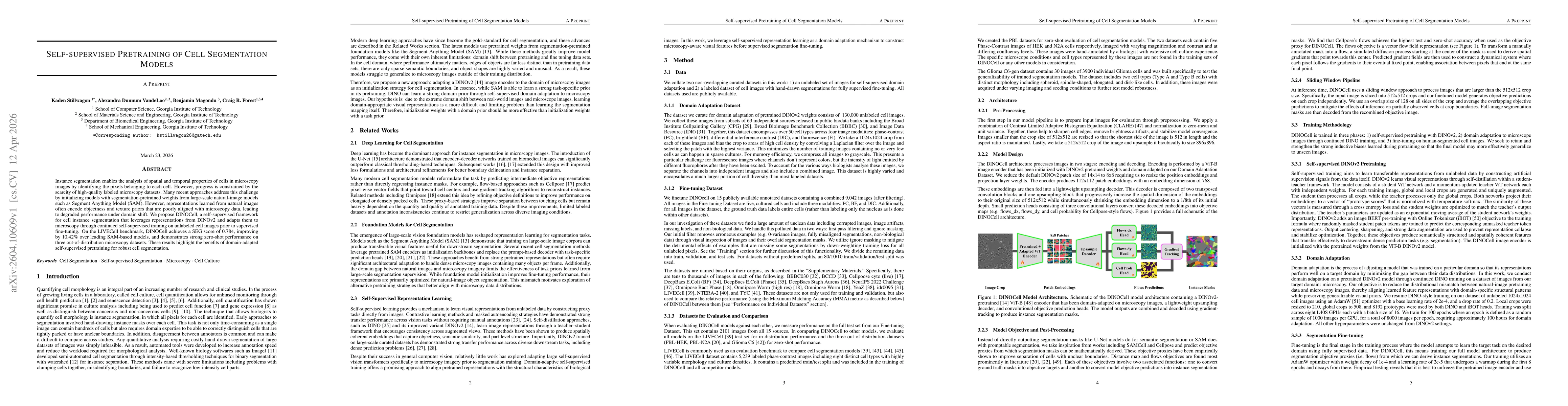
Instance segmentation enables the analysis of spatial and temporal properties of cells in microscopy images by identifying the pixels belonging to each cell. However, progress is constrained by the scarcity of high-quality labeled microscopy datasets. Many recent approaches address this challenge by initializing models with segmentation-pretrained weights from large-scale natural-image models such as Segment Anything Model (SAM). However, representations learned from natural images often encode objectness and texture priors that are poorly aligned with microscopy data, leading to degraded performance under domain shift. We propose DINOCell, a self-supervised framework for cell instance segmentation that leverages representations from DINOv2 and adapts them to microscopy through continued self-supervised training on unlabeled cell images prior to supervised fine-tuning. On the LIVECell benchmark, DINOCell achieves a SEG score of 0.784, improving by 10.42% over leading SAM-based models, and demonstrates strong zero-shot performance on three out-of-distribution microscopy datasets. These results highlight the benefits of domain-adapted self-supervised pretraining for robust cell segmentation.
AI Key Findings
Generated Apr 15, 2026
Methodology — What approach did the authors take?
Self-supervised pretraining of a ViT encoder with DINOv2, followed by domain adaptation via continued self-supervised training on unlabeled microscopy images, and final supervised fine-tuning on labeled cell segmentation data.
Key Results — What are the main findings?
- DINOCell achieves a SEG score of 0.784 on the LIVECell benchmark, a 10.42% improvement over SAM-based models.
- Pretraining boosts MMA by 29.51% on the fine-tuning test set and up to 285.49% on zero-shot datasets.
- PCA visualizations show that encoder representations become more aligned with cellular morphology after pretraining and domain adaptation.
Significance — Why does this research matter?
The study demonstrates that large-scale self-supervised pretraining can substantially improve cell segmentation performance and generalization to unseen microscopy domains, reducing reliance on costly labeled data.
Technical Contribution — What is the technical contribution?
A pipeline that combines DINOv2 self‑supervised pretraining with domain‑specific adaptation for cell instance segmentation, yielding state‑of‑the‑art performance without heavy annotation effort.
Novelty — What is new about this work?
First application of DINOv2 self‑supervised learning to microscopy, showing that domain‑adapted pretraining outperforms conventional SAM‑based initialization and provides robust zero‑shot generalization.
Limitations — What are the limitations of this study?
- Performance drops on highly dense or irregular datasets such as TYC, NTERA-2, and OmniposeWorm high‑res.
- Domain adaptation provides limited additional benefit after pretraining, suggesting diminishing returns.
Future Work — What did the authors propose for future work?
- Investigate stronger domain adaptation strategies or multi‑domain self‑supervised training to further improve out‑of‑distribution accuracy.
- Extend evaluation to more microscopy modalities and explore integration with other self‑supervised backbones.
Paper Details
How to Cite This Paper
@article{vandeloo2026self,
title = {Self-supervised Pretraining of Cell Segmentation Models},
author = {VandeLoo, Alexandra Dunnum and Magondu, Benjamin and Forest, Craig R. and others},
year = {2026},
eprint = {2604.10609},
archivePrefix = {arXiv},
primaryClass = {cs.CV},
}
VandeLoo, A., Magondu, B., Forest, C., & Stillwagon, K. (2026). Self-supervised Pretraining of Cell Segmentation Models. arXiv. https://arxiv.org/abs/2604.10609
VandeLoo, Alexandra Dunnum, et al. "Self-supervised Pretraining of Cell Segmentation Models." arXiv, 2026, arxiv.org/abs/2604.10609.
PDF Preview
Similar Papers
Found 4 papersSelf-Supervised Pretraining Improves Self-Supervised Pretraining
Devin Guillory, Colorado J. Reed, Xiangyu Yue et al.
Self-Supervised Pretraining for 2D Medical Image Segmentation
András Kalapos, Bálint Gyires-Tóth
Self-supervised Pretraining for Cardiovascular Magnetic Resonance Cine Segmentation
Rob A. J. de Mooij, Cian M. Scannell, Josien P. W. Pluim
Energy-Inspired Self-Supervised Pretraining for Vision Models
Jiang Wang, Zicheng Liu, Ze Wang et al.
Comments (0)