Authors
Publication
Metrics
Quick Actions
Quick Answers
What methodology did the authors use?
The study employed a semi-automated image analysis approach using machine learning segmentation and morphological thinning to analyze cellulose nanofibrils (CNFs). This involved training a segmentation model with Weka to identify CNF fibrils and background regions, followed by morphological thinning to generate skeleton segments for width and branching analysis. More in Methodology →
What are the key results?
The proposed method effectively differentiates between branching and network structures in CNFs, enabling accurate width and branching point analysis. — The skeleton pixel count is influenced by the aspect ratio and orientation of CNF fibrils, with shorter fibrils contributing more to skeleton counts when aspect ratios are low. More in Key Results →
Why is this work significant?
This research provides a robust framework for analyzing CNF morphology, which is critical for optimizing material properties in nanocellulose-based applications. The method enables precise characterization of fibril structure, supporting advancements in papermaking, biocomposites, and other nanocellulose applications. More in Significance →
What are the main limitations?
The method's accuracy is reduced when analyzing objects with aspect ratios less than 10, which may limit its applicability to certain CNF structures. — The segmentation performance depends on the quality and quantity of training data, which may require manual refinement for complex images. More in Limitations →
Paper Preview
Abstract
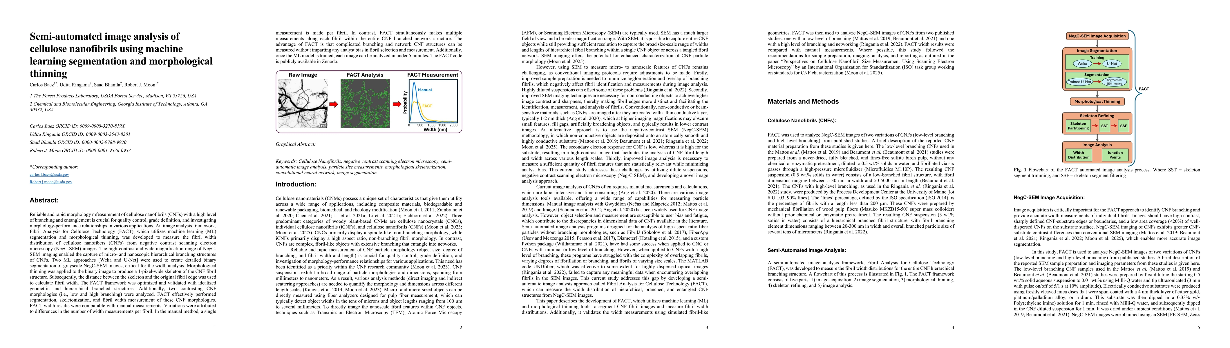
Reliable and rapid morphology measurement of cellulose nanofibrils (CNFs) with a high level of branching and entanglement is crucial for quality control, grade definition, and investigating morphology-performance relationships in various applications. An image analysis framework, Fibril Analysis for Cellulose Technology (FACT), which utilizes machine learning (ML) segmentation and morphological thinning, was developed to measure the fibril width distribution of cellulose nanofibers (CNFs) from negative contrast scanning electron microscopy (NegC-SEM) images. The high-contrast and wide magnification range of NegC-SEM imaging enabled the capture of micro- and nanoscopic hierarchical branching structures of CNFs. Two ML approaches [Weka and U-Net] were used to create detailed binary segmentation of grayscale NegC-SEM images, critical for the width analysis. Morphological thinning was applied to the binary image to produce a 1-pixel-wide skeleton of the CNF fibril structure. Subsequently, the distance between the skeleton and the original fibril edge was used to calculate fibril width. The FACT framework was optimized and validated with idealized geometric and hierarchical branched structures. FACT effectively performed segmentation, skeletonization, and fibril width measurement of these CNF morphologies. FACT width results were comparable with manual measurements. In the manual method, a single measurement is made per fibril. In contrast, FACT simultaneously makes multiple measurements along each fibril within the entire CNF branched network structure. The advantage of FACT is that complicated branching and network CNF structures can be measured without imparting any analyst bias in fibril selection and measurement. Additionally, once the ML model is trained, each image can be analyzed in under 5 minutes.
AI Key Findings
Generated Nov 17, 2025
Methodology — What approach did the authors take?
The study employed a semi-automated image analysis approach using machine learning segmentation and morphological thinning to analyze cellulose nanofibrils (CNFs). This involved training a segmentation model with Weka to identify CNF fibrils and background regions, followed by morphological thinning to generate skeleton segments for width and branching analysis.
Key Results — What are the main findings?
- The proposed method effectively differentiates between branching and network structures in CNFs, enabling accurate width and branching point analysis.
- The skeleton pixel count is influenced by the aspect ratio and orientation of CNF fibrils, with shorter fibrils contributing more to skeleton counts when aspect ratios are low.
- The method demonstrated improved statistical analysis of CNF widths when most objects have aspect ratios greater than 10.
Significance — Why does this research matter?
This research provides a robust framework for analyzing CNF morphology, which is critical for optimizing material properties in nanocellulose-based applications. The method enables precise characterization of fibril structure, supporting advancements in papermaking, biocomposites, and other nanocellulose applications.
Technical Contribution — What is the technical contribution?
The technical contribution lies in the integration of machine learning segmentation with morphological thinning to enable precise and scalable analysis of CNF morphology, including branching and network structures.
Novelty — What is new about this work?
This work introduces a novel semi-automated approach combining machine learning and morphological thinning for CNF analysis, offering improved accuracy and efficiency compared to traditional manual or semi-automated methods.
Limitations — What are the limitations of this study?
- The method's accuracy is reduced when analyzing objects with aspect ratios less than 10, which may limit its applicability to certain CNF structures.
- The segmentation performance depends on the quality and quantity of training data, which may require manual refinement for complex images.
Future Work — What did the authors propose for future work?
- Developing adaptive algorithms to handle objects with varying aspect ratios and edge shapes.
- Integrating real-time feedback mechanisms to improve segmentation accuracy in complex or noisy images.
- Expanding the method to analyze three-dimensional CNF structures using advanced imaging techniques.
How to Cite This Paper
@article{baez2025semi,
  title         = {Semi-automated image analysis of Cellulose Nanofibrils using Machine
  learning segmentation and Morphological thinning},
  author        = {Baez, Carlos and Moon, Robert J. and Bhamla, Saad and others},
  year          = {2025},
  eprint        = {2509.06618},
  archivePrefix = {arXiv},
  primaryClass  = {physics.bio-ph},
}Baez, C., Moon, R., Bhamla, S., & Ringania, U. (2025). Semi-automated image analysis of Cellulose Nanofibrils using Machine
learning segmentation and Morphological thinning. arXiv. https://arxiv.org/abs/2509.06618Baez, Carlos, et al. "Semi-automated image analysis of Cellulose Nanofibrils using Machine
learning segmentation and Morphological thinning." arXiv, 2025, arxiv.org/abs/2509.06618.PDF Preview
Similar Papers
Found 4 papersDynamic characterization of cellulose nanofibrils in sheared and extended semi-dilute dispersions
Nitesh Mittal, L. Daniel Söderberg, Fredrik Lundell et al.
Segmentation and Analysis of a Sketched Truss Frame Using Morphological Image Processing Techniques
Oguz Gunes, Mirsalar Kamari
Semi-automated detection of cleaning interactions using supervised machine learning.
Paula, José Ricardo, Garcia, Nuno Cruz, Oliveira, Raul
Comments (0)