Semi-supervised variational autoencoder for cell feature extraction in multiplexed immunofluorescence images

Source: ArXiv

AI Quick Summary

The paper proposes a semi-supervised variational autoencoder to extract complex cell features from multiplexed immunofluorescence images, aiming to improve cell phenotype classification over existing methods. The model is validated using over 44,000 image patches from breast cancer tissue microarrays.

Share: Share on X LinkedIn

Summary

Advancements in digital imaging technologies have sparked increased interest in using multiplexed immunofluorescence (mIF) images to visualise and identify the interactions between specific immunophenotypes with the tumour microenvironment at the cellular level. Current state-of-the-art multiplexed immunofluorescence image analysis pipelines depend on cell feature representations characterised by morphological and stain intensity-based metrics generated using simple statistical and machine learning-based tools. However, these methods are not capable of generating complex representations of cells. We propose a deep learning-based cell feature extraction model using a variational autoencoder with supervision using a latent subspace to extract cell features in mIF images. We perform cell phenotype classification using a cohort of more than 44,000 multiplexed immunofluorescence cell image patches extracted across 1,093 tissue microarray cores of breast cancer patients, to demonstrate the success of our model against current and alternative methods.

AI Key Findings

Generated Sep 03, 2025

Methodology

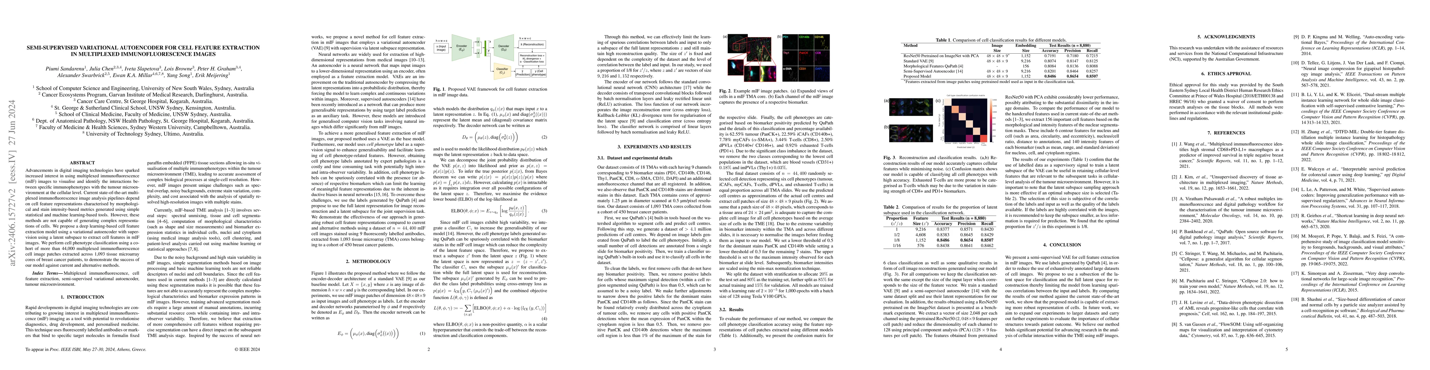
The research proposes a semi-supervised variational autoencoder (VAE) for cell feature extraction in multiplexed immunofluorescence (mIF) images. The method uses a latent subspace for supervision to overcome limitations of traditional methods in capturing complex cell representations. It integrates a classifier to enhance generalization and limits learning of spurious correlations between labels and inputs to a subspace of full latent representations.

Key Results

  • The proposed model outperformed current and alternative methods in cell phenotype classification, achieving an accuracy of 0.8486 on a dataset of 44,400 mIF cell image patches.
  • The model demonstrated robust cell feature representations, with high reconstruction quality while limiting spurious correlations between labels and inputs.
  • The study found that an optimal proportion of z′/z in the latent space for classification was 1/8, enhancing the retention of cellular-level features relevant to subsequent tasks in tumour microenvironment analysis.

Significance

This research is significant as it advances cellular-level analysis of the tumour microenvironment using mIF images, offering a more accurate and robust cell feature extraction method compared to existing techniques.

Technical Contribution

The main technical contribution is the development of a semi-supervised VAE for cell feature extraction in mIF images, which effectively uses a latent subspace for supervision and integrates a classifier to enhance generalization.

Novelty

The novelty of this work lies in its application of a semi-supervised VAE tailored for complex cell feature extraction in mIF images, addressing limitations of traditional methods and outperforming current alternatives in cell phenotype classification.

Limitations

  • The study relied on cell phenotype labels generated by QuPath, which could be subject to inter- and intra-observer variability.
  • The performance of the model might be affected by the significant class imbalance in the dataset, requiring careful handling of class labels.

Future Work

  • Expand experiments to larger datasets for further validation and evaluation.
  • Investigate the importance of cellular structures in relation to patient outcomes.

Paper Details

PDF Preview

Download PDF

Key Terms

cell (0.303)
learning based (0.218)
representations (0.180)
feature (0.163)
images (0.162)
image (0.159)
advancements (0.151)
patches (0.149)

Citation Network

Current paper (gray), citations (green), references (blue)

Display is limited for performance on very large graphs.

Current Paper
Citations
References
Click to view

No citations found for this paper.

Comments (0)

This website uses cookies to ensure you get the best experience. Learn more