Separate-scan atomic force microscope for fast infrared scattering-type scanning near-field optical microscope
Publication
Metrics
Paper Preview
Abstract
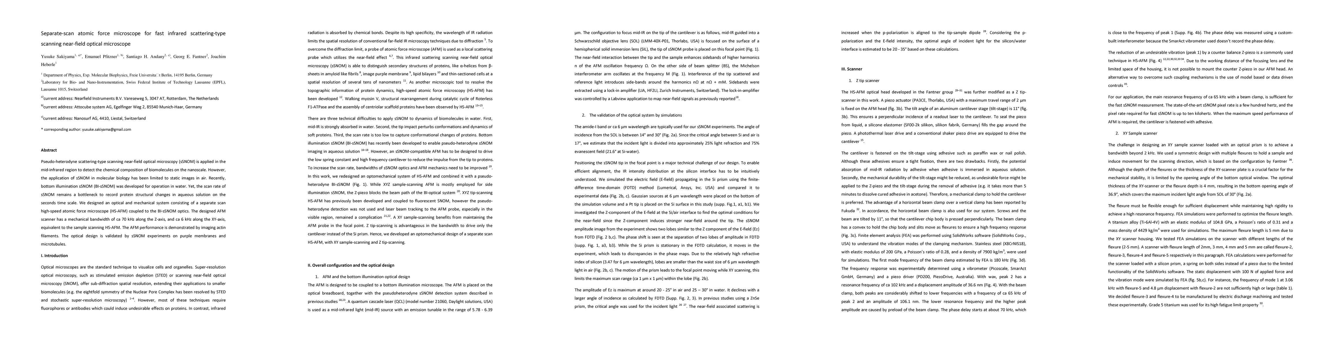
Pseudo-heterodyne scattering-type scanning near-field optical microscopy (sSNOM) is applied in the mid-infrared region to detect the chemical composition of biomolecules on the nanoscale. However, the application of sSNOM in molecular biology has been limited to static images in air. Recently, bottom illumination sSNOM (BI-sSNOM) was developed for operation in water. Yet, the scan rate of sSNOM remains a bottleneck to record protein structural changes in aqueous solution on the seconds time scale. We designed an optical and mechanical system consisting of a separate scan high-speed atomic force microscope (HS-AFM) coupled to the BI-sSNOM optics. The designed AFM scanner has a mechanical bandwidth of ca 70 kHz along the Z-axis, and ca 6 kHz along the XY-axis, equivalent to the sample scanning HS-AFM. The AFM performance is demonstrated by imaging actin filaments. The optical design is validated by sSNOM experiments on purple membranes and microtubules.
AI Key Findings
Get AI-generated insights about this paper's methodology, results, significance, and more — seven facets brought into focus.
Discussion 0