Quick Summary:

A new method called SepaReg improves automatic delineation of organs-at-risk and gross-tumor-volume in radiotherapy planning by learning to separate regions within images, achieving better performance than existing state-of-the-art approaches.

Summary

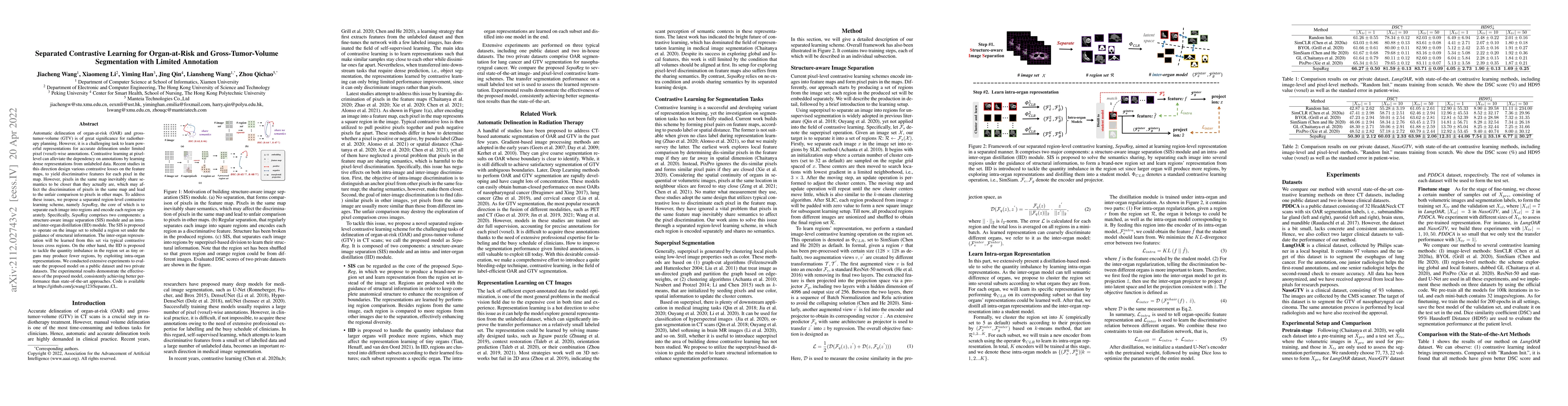
Automatic delineation of organ-at-risk (OAR) and gross-tumor-volume (GTV) is of great significance for radiotherapy planning. However, it is a challenging task to learn powerful representations for accurate delineation under limited pixel (voxel)-wise annotations. Contrastive learning at pixel-level can alleviate the dependency on annotations by learning dense representations from unlabeled data. Recent studies in this direction design various contrastive losses on the feature maps, to yield discriminative features for each pixel in the map. However, pixels in the same map inevitably share semantics to be closer than they actually are, which may affect the discrimination of pixels in the same map and lead to the unfair comparison to pixels in other maps. To address these issues, we propose a separated region-level contrastive learning scheme, namely SepaReg, the core of which is to separate each image into regions and encode each region separately. Specifically, SepaReg comprises two components: a structure-aware image separation (SIS) module and an intra- and inter-organ distillation (IID) module. The SIS is proposed to operate on the image set to rebuild a region set under the guidance of structural information. The inter-organ representation will be learned from this set via typical contrastive losses cross regions. On the other hand, the IID is proposed to tackle the quantity imbalance in the region set as tiny organs may produce fewer regions, by exploiting intra-organ representations. We conducted extensive experiments to evaluate the proposed model on a public dataset and two private datasets. The experimental results demonstrate the effectiveness of the proposed model, consistently achieving better performance than state-of-the-art approaches. Code is available at https://github.com/jcwang123/Separate_CL.

Paper Details

PDF Preview

Download PDF

Key Terms

contrastive (0.241)
contrastive (0.239)
pixels (0.206)
pixel (0.195)
pixel (0.195)
annotations (0.168)
annotations (0.166)
losses (0.160)
intra (0.157)
proposed model (0.154)
intra (0.152)
losses (0.151)
representations (0.148)
region (0.148)
map (0.148)
map (0.147)
proposed model (0.147)
region (0.144)
representations (0.143)
regions (0.140)

Citation Network

Current paper (gray), citations (green), references (blue)

Current Paper
Citations
References
Click to view
Title Authors Year Actions
Multi-view cross-consistency and multi-scale cross-layer contrastive learning for semi-supervised medical image segmentation
Xunhang Cao, Xiaoxin Guo, Guangqi Yang, Qi Chen, Hongliang Dong
2025
View Paper
WT-BCP: Wavelet Transform based Bidirectional Copy-Paste for Semi-Supervised Medical Image Segmentation
Mingya Zhang, Liang Wang, Limei Gu, Tingshen Ling, Xianping Tao
2025
View Paper
SP3: Superpixel-propagated pseudo-label learning for weakly semi-supervised medical image segmentation
Shiman Li, Jiayue Zhao, Shaolei Liu, Xiaokun Dai, Chenxi Zhang, Zhijian Song
2024
View Paper
Data augmentation strategies for semi-supervised medical image segmentation
Jiahui Wang, Dongshen Ruan, Yang Li, Zefeng Wang, Yongquan Wu, Tao Tan, Guang Yang, Mingfeng Jiang
2024
View Paper
Deep semi-supervised learning for medical image segmentation: A review
Kai Han, Victor S. Sheng, Yuqing Song, Yi Liu, Chengjian Qiu, Siqi Ma, Zhe Liu
2024
View Paper
Review of Semi-Supervised Medical Image Segmentation based on the U-Net
Lina Dai, M. Johar, M. H. Alkawaz
2024
View Paper
OpenESS: Event-Based Semantic Scene Understanding with Open Vocabularies
Lingdong Kong, You-Chen Liu, Lai Xing Ng, Benoit R. Cottereau, Wei Tsang Ooi
2024
View Paper
Constructing and Exploring Intermediate Domains in Mixed Domain Semi-supervised Medical Image Segmentation
Qinghe Ma, Jian Zhang, Lei Qi, Qian Yu, Yinghuan Shi, Yang Gao
2024
View Paper
DCL-Net: Dual Contrastive Learning Network for Semi-Supervised Multi-Organ Segmentation
L. Wen, Zheng-Kai Feng, Yun Hou, Peng Wang, Xi Wu, Jiliu Zhou, Yan Wang
2024
View Paper
SC-SSL: Self-Correcting Collaborative and Contrastive Co-Training Model for Semi-Supervised Medical Image Segmentation
Juzheng Miao, Si-Ping Zhou, Guangquan Zhou, Kaini Wang, Meng Yang, Shoujun Zhou, Yang Chen
2023
View Paper
Entropy-guided contrastive learning for semi-supervised medical image segmentation
Junsong Xie, Qian Wu, Renju Zhu
2023
View Paper
Overcoming the Challenges in the Development and Implementation of Artificial Intelligence in Radiology: A Comprehensive Review of Solutions Beyond Supervised Learning
G. Hong, Miso Jang, Sunggu Kyung, Kyungjin Cho, Jiheon Jeong, Grace Yoojin Lee, Keewon Shin, Ki Duk Kim, Seung Min Ryu, J. Seo, Sang Min Lee, Namkug Kim
2023
View Paper
Bidirectional Copy-Paste for Semi-Supervised Medical Image Segmentation
Yunhao Bai, Duowen Chen, Qingli Li, Wei-lei Shen, Yan Wang
2023
View Paper
Multi-organ segmentation: a progressive exploration of learning paradigms under scarce annotation
Shiman Li, Haoran Wang, Yucong Meng, Chenxi Zhang, Zhijian Song
2023
View Paper
Learning with Limited Annotations: A Survey on Deep Semi-Supervised Learning for Medical Image Segmentation
Rushi Jiao, Yichi Zhang, Leiting Ding, Rong Cai, Jicong Zhang
2022
View Paper
Segmentation of teeth restoration from X-ray bitewing images
David Grundfest
2023
View Paper

Comments (0)

This website uses cookies to ensure you get the best experience. Learn more