Summary
Overlap-based metrics such as the Dice Similarity Coefficient (DSC) penalize segmentation errors more heavily in smaller structures. As organ size differs by sex, this implies that a segmentation error of equal magnitude may result in lower DSCs in women due to their smaller average organ volumes compared to men. While previous work has examined sex-based differences in models or datasets, no study has yet investigated the potential bias introduced by the DSC itself. This study quantifies sex-based differences of the DSC and the normalized DSC in an idealized setting independent of specific models. We applied equally-sized synthetic errors to manual MRI annotations from 50 participants to ensure sex-based comparability. Even minimal errors (e.g., a 1 mm boundary shift) produced systematic DSC differences between sexes. For small structures, average DSC differences were around 0.03; for medium-sized structures around 0.01. Only large structures (i.e., lungs and liver) were mostly unaffected, with sex-based DSC differences close to zero. These findings underline that fairness studies using the DSC as an evaluation metric should not expect identical scores between men and women, as the metric itself introduces bias. A segmentation model may perform equally well across sexes in terms of error magnitude, even if observed DSC values suggest otherwise. Importantly, our work raises awareness of a previously underexplored source of sex-based differences in segmentation performance. One that arises not from model behavior, but from the metric itself. Recognizing this factor is essential for more accurate and fair evaluations in medical image analysis.
AI Key Findings
Generated Sep 28, 2025
Methodology
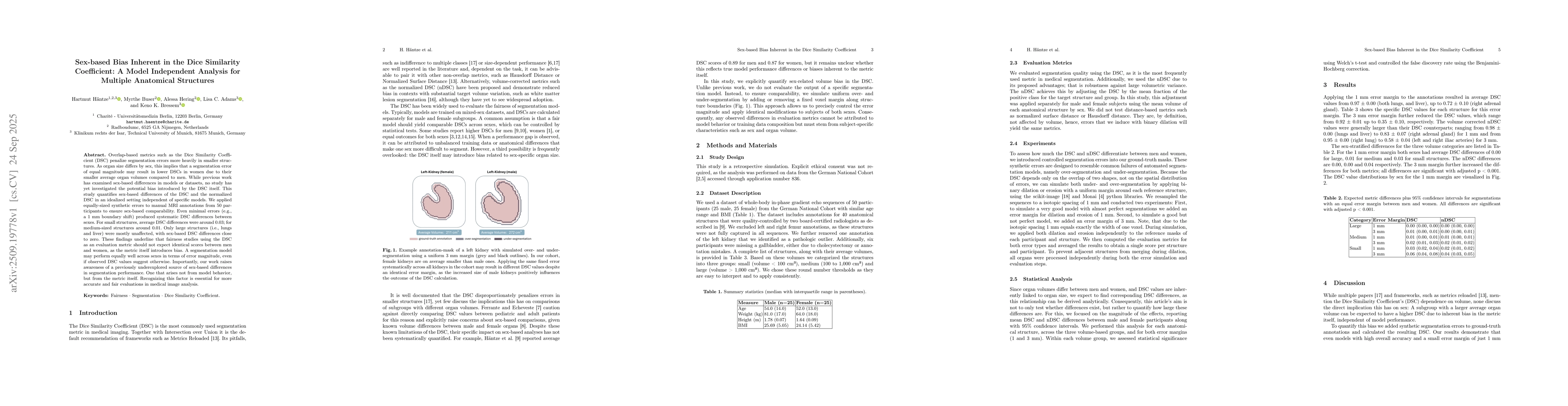
The study introduced controlled segmentation errors into synthetic MRI annotations from 50 participants, applying binary dilation/erosion with uniform margins to simulate over/under-segmentation. They analyzed DSC and normalized DSC differences across anatomical structures categorized by size (small, medium, large) and evaluated statistical significance using Welch's t-test with Benjamini-Hochberg correction.
Key Results
- Sex-based DSC differences were significant for small structures (avg 0.03), medium (0.01), but negligible for large structures (close to 0).
- Normalized DSC (nDSC) showed reduced sex-based differences compared to standard DSC, especially for small structures.
- Even minimal errors (1mm margin) produced systematic DSC differences between sexes, indicating inherent metric bias.
Significance
This research highlights that the Dice Similarity Coefficient (DSC) itself introduces sex-based bias due to anatomical volume differences, which is critical for fair evaluation in medical image segmentation. It warns against interpreting DSC disparities as model bias when they may stem from the metric's design.
Technical Contribution
Quantified the inherent sex-based bias in DSC through controlled synthetic error simulations, demonstrating how volume differences affect metric outcomes independently of model performance.
Novelty
First study to isolate and quantify DSC's inherent sex-based bias, showing that metric design, not model behavior, can cause systematic differences in segmentation evaluation between sexes.
Limitations
- Results are based on a German cohort, limiting generalizability to other populations.
- Simulated errors via binary dilation/erosion may not fully capture real-world segmentation complexities.
Future Work
- Investigate DSC bias in other imaging modalities like CT or 2D X-ray.
- Develop alternative metrics less sensitive to volume-based sex differences.
- Explore how these findings impact clinical decision-making and model fairness assessments.
Paper Details
PDF Preview
Similar Papers
Found 4 papersTackling Bias in the Dice Similarity Coefficient: Introducing nDSC for White Matter Lesion Segmentation
Nataliia Molchanova, Meritxell Bach Cuadra, Mark Gales et al.
Theoretical analysis and experimental validation of volume bias of soft Dice optimized segmentation maps in the context of inherent uncertainty
David Robben, Jeroen Bertels, Dirk Vandermeulen et al.
TotalSegmentator MRI: Sequence-Independent Segmentation of 59 Anatomical Structures in MR images
Thomas Küstner, Shan Yang, Matthias Jung et al.
Radiomics as a measure superior to the Dice similarity coefficient for tumor segmentation performance evaluation
Yoichi Watanabe, Rukhsora Akramova
Comments (0)