Publication
Metrics
Quick Actions
AI Quick Summary
This paper uses CycleGAN with shape-aware loss functions to generate synthetic pathological lung CT scans, addressing the challenge of segmenting peripheral opacities. It introduces a novel L1 loss to maintain shape consistency and refines inputs with preprocessing, improving semi-supervised lung segmentation via U-Net models trained on augmented data.
Paper Preview
Abstract
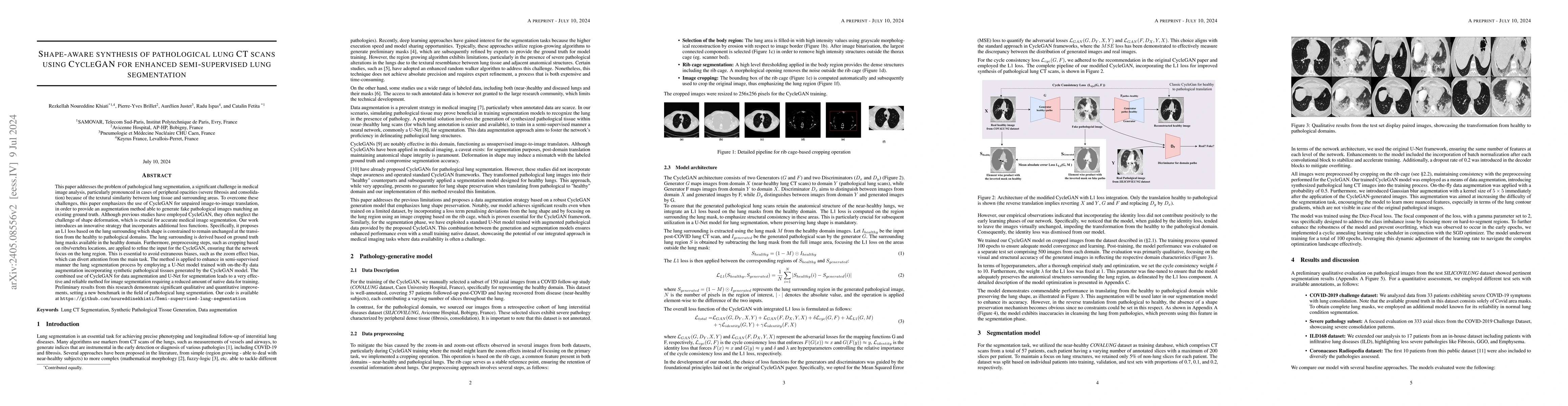
This paper addresses the problem of pathological lung segmentation, a significant challenge in medical image analysis, particularly pronounced in cases of peripheral opacities (severe fibrosis and consolidation) because of the textural similarity between lung tissue and surrounding areas. To overcome these challenges, this paper emphasizes the use of CycleGAN for unpaired image-to-image translation, in order to provide an augmentation method able to generate fake pathological images matching an existing ground truth. Although previous studies have employed CycleGAN, they often neglect the challenge of shape deformation, which is crucial for accurate medical image segmentation. Our work introduces an innovative strategy that incorporates additional loss functions. Specifically, it proposes an L1 loss based on the lung surrounding which shape is constrained to remain unchanged at the transition from the healthy to pathological domains. The lung surrounding is derived based on ground truth lung masks available in the healthy domain. Furthermore, preprocessing steps, such as cropping based on ribs/vertebra locations, are applied to refine the input for the CycleGAN, ensuring that the network focus on the lung region. This is essential to avoid extraneous biases, such as the zoom effect bias, which can divert attention from the main task. The method is applied to enhance in semi-supervised manner the lung segmentation process by employing a U-Net model trained with on-the-fly data augmentation incorporating synthetic pathological tissues generated by the CycleGAN model. Preliminary results from this research demonstrate significant qualitative and quantitative improvements, setting a new benchmark in the field of pathological lung segmentation. Our code is available at https://github.com/noureddinekhiati/Semi-supervised-lung-segmentation
AI Key Findings
Get AI-generated insights about this paper's methodology, results, and significance.
How to Cite This Paper
@article{justet2024shape,
  title         = {Shape-aware synthesis of pathological lung CT scans using CycleGAN for
  enhanced semi-supervised lung segmentation},
  author        = {Justet, Aurélien and Fetita, Catalin and Brillet, Pierre-Yves and others},
  year          = {2024},
  eprint        = {2405.08556},
  archivePrefix = {arXiv},
  primaryClass  = {eess.IV},
}Justet, A., Fetita, C., Brillet, P., Ispas, R., & Khiati, R. (2024). Shape-aware synthesis of pathological lung CT scans using CycleGAN for
enhanced semi-supervised lung segmentation. arXiv. https://arxiv.org/abs/2405.08556Justet, Aurélien, et al. "Shape-aware synthesis of pathological lung CT scans using CycleGAN for
enhanced semi-supervised lung segmentation." arXiv, 2024, arxiv.org/abs/2405.08556.PDF Preview
Key Terms
Citation Network
Current paper (gray), citations (green), references (blue)
Display is limited for performance on very large graphs.
Similar Papers
Found 4 papersDiff-Lung: Diffusion-Based Texture Synthesis for Enhanced Pathological Tissue Segmentation in Lung CT Scans
Rezkellah Noureddine Khiati, Pierre-Yves Brillet, Radu Ispas et al.
Enhanced detection of the presence and severity of COVID-19 from CT scans using lung segmentation
Robert Turnbull
Semi-Supervised Segmentation of Radiation-Induced Pulmonary Fibrosis from Lung CT Scans with Multi-Scale Guided Dense Attention
Byong Yi, Guotai Wang, Shuwei Zhai et al.
| Title | Authors | Year | Actions |
|---|
Comments (0)