This paper proposes a shape-constrained CNN for cardiac MR segmentation that integrates a statistical shape model to predict realistic left ventricle and myocardial shapes, while simultaneously estimating pose and shape parameters. The method achieves high correlation in segmentation metrics and state-of-the-art results on both in-house and public datasets.
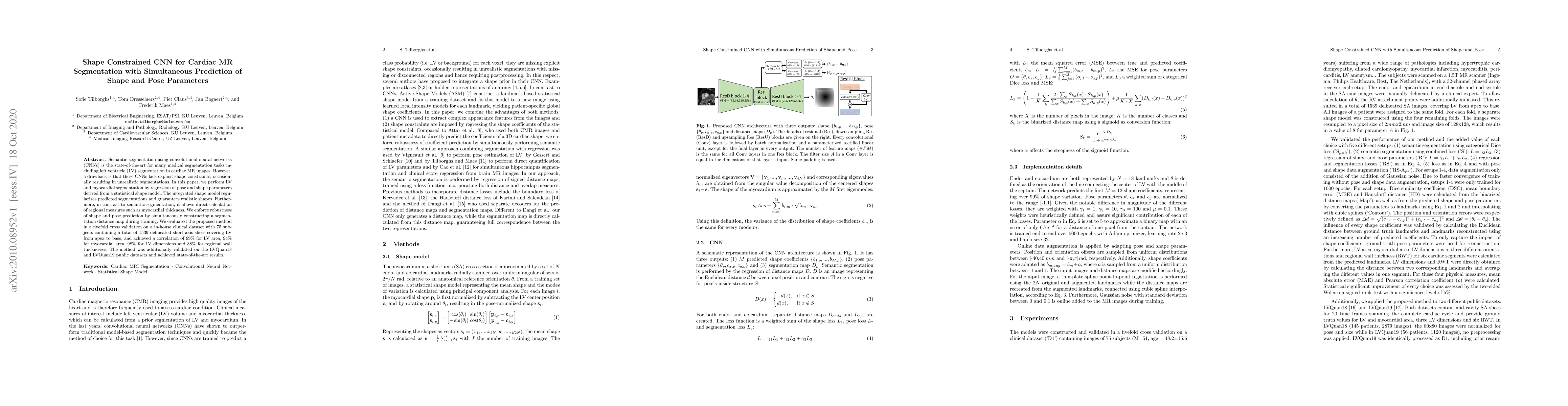
Semantic segmentation using convolutional neural networks (CNNs) is the state-of-the-art for many medical segmentation tasks including left ventricle (LV) segmentation in cardiac MR images. However, a drawback is that these CNNs lack explicit shape constraints, occasionally resulting in unrealistic segmentations. In this paper, we perform LV and myocardial segmentation by regression of pose and shape parameters derived from a statistical shape model. The integrated shape model regularizes predicted segmentations and guarantees realistic shapes. Furthermore, in contrast to semantic segmentation, it allows direct calculation of regional measures such as myocardial thickness. We enforce robustness of shape and pose prediction by simultaneously constructing a segmentation distance map during training. We evaluated the proposed method in a fivefold cross validation on a in-house clinical dataset with 75 subjects containing a total of 1539 delineated short-axis slices covering LV from apex to base, and achieved a correlation of 99% for LV area, 94% for myocardial area, 98% for LV dimensions and 88% for regional wall thicknesses. The method was additionally validated on the LVQuan18 and LVQuan19 public datasets and achieved state-of-the-art results.
This graph shows how this paper is connected to other research. The current paper is shown in dark gray, papers that cite it are in green, and papers it references are in blue. You can:
• Hover over any node to see the paper title
• Click a node to view that paper's details
• Drag nodes to rearrange the graph
• Use the zoom controls (bottom right) to zoom in/out
Title | Authors | Year | Actions |
---|---|---|---|
Simultaneous Hip Implant Segmentation and Gruen Landmarks Detection
|
Asma Alzaid, B. Lineham, S. Dogramadzi, H. Pandit, A. Frangi, S. Xie
|
2023
|
View Paper |
Automated Diagnosis of Cardiovascular Diseases from Cardiac Magnetic Resonance Imaging Using Deep Learning Models: A Review
|
M. Jafari, A. Shoeibi, Marjane Khodatars, Navid Ghassemi, Parisa Moridian, Niloufar Delfan, R. Alizadehsani, Abbas Khosravi, Saiguang Ling, Yu-Dong Zhang, Shui-Hua Wang, J. M. Górriz, Hamid Alinejad-Rokny, U. R. Acharya
|
2022
|
View Paper |
Deep Statistic Shape Model for Myocardium Segmentation
|
Xiaoling Hu, Xiao Chen, Yikang Liu, Eric Z. Chen, Terrence Chen, Shanhui Sun
|
2022
|
View Paper |
Peripapillary Atrophy Segmentation Based on ASM Loss
|
Mengxuan Li, He Zhao, Jie Xu, Huiqi Li
|
2022
|
View Paper |
Shape constrained CNN for segmentation guided prediction of myocardial shape and pose parameters in cardiac MRI
|
Sofie Tilborghs, J. Bogaert, F. Maes
|
2022
|
View Paper |
Medical Image Segmentation With Limited Supervision: A Review of Deep Network Models
|
Jialin Peng, Ye Wang
|
2021
|
View Paper |
A Survey on Shape-Constraint Deep Learning for Medical Image Segmentation
|
Simon Bohlender, Ilkay Öksüz, A. Mukhopadhyay
|
2021
|
View Paper |
Atlas-ISTN: Joint Segmentation, Registration and Atlas Construction with Image-and-Spatial Transformer Networks
|
Matthew Sinclair, A. Schuh, K. Hahn, K. Petersen, Ying Bai, James Batten, M. Schaap, B. Glocker
|
2020
|
View Paper |
Slice-Mask Based 3D Cardiac Shape Reconstruction from CT Volume
|
Xiaohan Yuan, Cong Liu, Fu Feng, Yinsu Zhu, Yangang Wang
|
2022
|
View Paper |
Shape Feature Loss for Kidney Segmentation in 3D Ultrasound Images
|
Haithem Boussaid, L. Rouet
|
2021
|
View Paper |
Frederik Maes, Sofie Tilborghs, Jan Bogaert
Published: 2022-08-18
Published: 2017-09-14
Published: 2019-12-18
Comments (0)