ArxivLens

Quick Summary:

The paper proposes a sharpness loss regularized GAN to synthesize realistic histopathology images with clear nuclei contours, addressing the issue of generating clear contours for overlapped and touching nuclei. The method uses a normalized nucleus distance map to encode nuclei contour information and is evaluated using four image quality metrics and segmentation results, showing superior performance.

arXiv Id: 2110.14709
Date Published: 2021-10-29
Date Updated: 2021-10-29

Categories:

Key Terms (TF-IDF):

nuclei (0.269)
nuclei (0.262)
generative adversarial (0.227)
images (0.212)
images (0.211)
adversarial (0.198)
generate (0.196)
generative (0.192)
generate (0.192)
generative (0.191)
adversarial (0.189)
clear (0.183)
clear (0.179)
realistic (0.175)
realistic (0.171)
loss (0.158)
loss (0.155)
proposed (0.151)
proposed (0.150)
image (0.147)
License: http://creativecommons.org/licenses/by-nc-nd/4.0/

Summary:

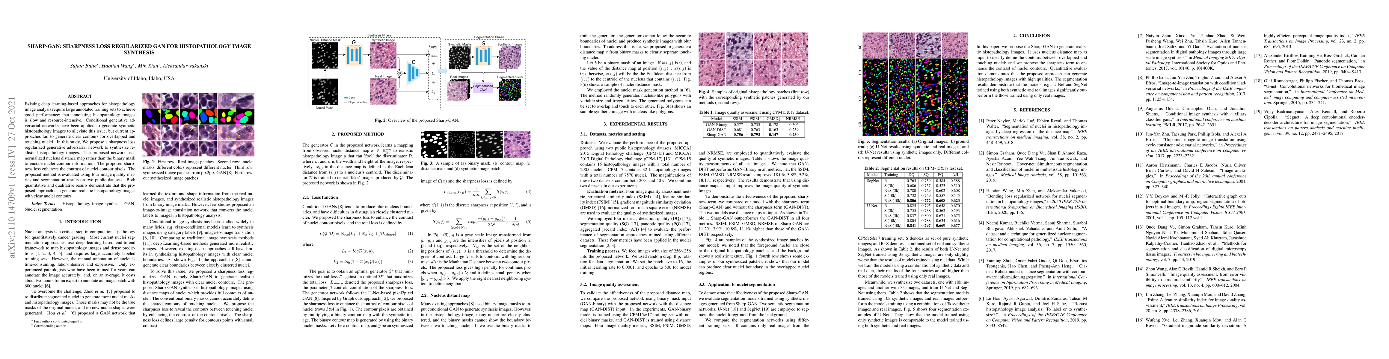
Existing deep learning-based approaches for histopathology image analysis require large annotated training sets to achieve good performance; but annotating histopathology images is slow and resource-intensive. Conditional generative adversarial networks have been applied to generate synthetic histopathology images to alleviate this issue, but current approaches fail to generate clear contours for overlapped and touching nuclei. In this study, We propose a sharpness loss regularized generative adversarial network to synthesize realistic histopathology images. The proposed network uses normalized nucleus distance map rather than the binary mask to encode nuclei contour information. The proposed sharpness loss enhances the contrast of nuclei contour pixels. The proposed method is evaluated using four image quality metrics and segmentation results on two public datasets. Both quantitative and qualitative results demonstrate that the proposed approach can generate realistic histopathology images with clear nuclei contours.

Citation & Reference Network Visualization

This graph shows how this paper is connected to other research. The current paper is shown in dark gray, papers that cite it are in green, and papers it references are in blue. You can:

Current Paper
Papers citing this work (Citations)
Papers cited by this work (References)

• Hover over any node to see the paper title

• Click a node to view that paper's details

• Drag nodes to rearrange the graph

• Use the zoom controls (bottom right) to zoom in/out

Citations and References

Title Authors Year Actions
Advancing Histopathology with Deep Learning Under Data Scarcity: A Decade in Review
Ahmad Obeid, Said Boumaraf, A. Sohail, Taimur Hassan, Sajid Javed, Jorge Dias, Mohammed Bennamoun, N. Werghi
2024
View Paper
Enhanced Generation of Gastrointestinal Images for Data Augmentation Using a Modified BlobGAN
Aula Ayubi, N. P. Utama
2024
View Paper
Co-synthesis of Histopathology Nuclei Image-Label Pairs using a Context-Conditioned Joint Diffusion Model
Seonghui Min, Hyunwoo Oh, Won-Ki Jeong
2024
View Paper
Method and Software Tool for Generating Artificial Databases of Biomedical Images Based on Deep Neural Networks
O. Berezsky, Petro B. Liashchynskyi, O. Pitsun, G. Melnyk
2024
View Paper
Photo-realistic photo synthesis using improved conditional generative adversarial networks
Raghavendra Shetty Mandara Kirimanjeshwara, S. N. Prasad
2024
View Paper
Nuclei Segmentation Method based on CycleGAN-generated Images
Jie Shu, Qing Wang, Shanlin Liu
2023
View Paper
Generative Adversarial Networks in Digital Histopathology: Current Applications, Limitations, Ethical Considerations, and Future Directions.
Shahd A. Alajaji, Zaid H. Khoury, Mohamed A. Elgharib, Mamoon Saeed, Ahmed R.H. Ahmed, Mohammad B. Khan, Tiffany Tavares, Maryam Jessri, A. Puche, Hamid Hoorfar, Ivan J Stojanov, James J. Sciubba, Ahmed S. Sultan
2023
View Paper
Microscopy Image Segmentation via Point and Shape Regularized Data Synthesis
Shijie Li, Mengwei Ren, T. Ach, G. Gerig
2023
View Paper
Panoptic quality should be avoided as a metric for assessing cell nuclei segmentation and classification in digital pathology
Adrien Foucart, O. Debeir, C. Decaestecker
2023
View Paper
Inferring Parameters of Pyramidal Neuron Excitability in Mouse Models of Alzheimer’s Disease Using Biophysical Modeling and Deep Learning
Soheil Saghafi, Timothy H. Rumbell, V. Gurev, J. Kozloski, Francesco Tamagnini, K. Wedgwood, C. Diekman
2023
View Paper
Topology-Guided Multi-Class Cell Context Generation for Digital Pathology
Shahira Abousamra, Rajarsi R. Gupta, T. Kurç, D. Samaras, J. Saltz, Chao Chen
2023
View Paper
Evaluating participating methods in image analysis challenges: Lessons from MoNuSAC 2020
Adrien Foucart, O. Debeir, C. Decaestecker
2023
View Paper
Enhanced Sharp-Gan for Histopathology Image Synthesis
S. Butte, Haotian Wang, Aleksandar Vakanski, Min Xian
2023
View Paper
SIAN: Style-Guided Instance-Adaptive Normalization for Multi-Organ Histopathology Image Synthesis
Haotian Wang, Min Xian, Aleksandar Vakanski, Bryar Shareef
2022
View Paper
NormToRaw: A Style Transfer Based Self-supervised Learning Approach for Nuclei Segmentation
Xianlai Chen, Xuantong Zhong, Taixiang Li, Ying An, L. Mo
2022
View Paper
Director’s Letter
Meme Omogbai
2020
View Paper
Knowledge Distillation Generative Adversarial Network for Image-to-Image Translation
C. Sub-r-pa, R. Chen
2024
View Paper

Similar Papers

Comments (0)

This website uses cookies to ensure you get the best experience. Learn more