Sigmoid function based intensity transformation for parameter initialization in MRI-PET Registration Tool for Preclinical Studies
Publication
Metrics
AI Quick Summary
Researchers developed a tool for registering MRI-PET images to fuse functional data with anatomical information, improving accuracy in preclinical studies. A two-stage registration approach uses principal component analysis and non-uniform intensity transformation to enhance low-intensity PET volumes.
Paper Preview
Abstract
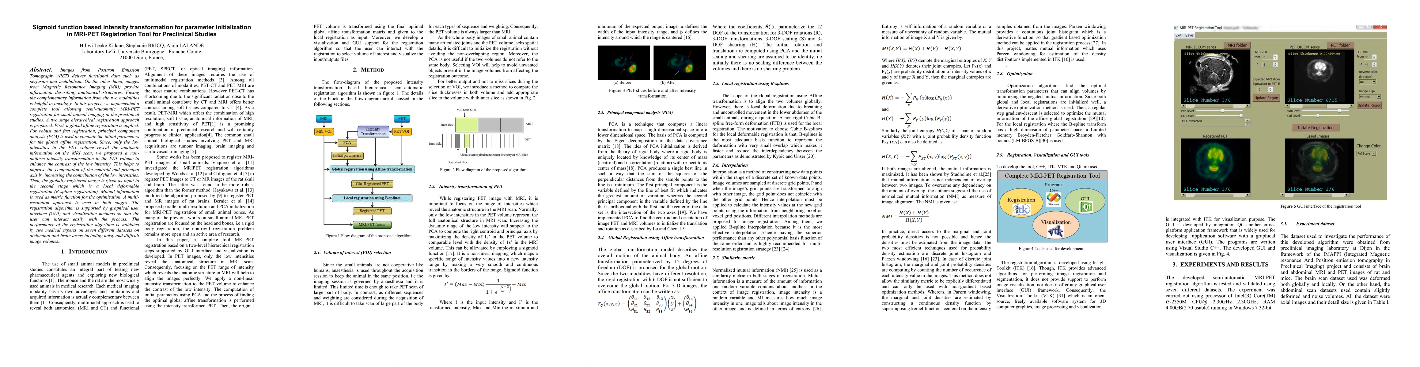
Images from Positron Emission Tomography (PET) deliver functional data such as perfusion and metabolism. On the other hand, images from Magnetic Resonance Imaging (MRI) provide information describing anatomical structures. Fusing the complementary information from the two modalities is helpful in oncology. In this project, we implemented a complete tool allowing semi-automatic MRI-PET registration for small animal imaging in the preclinical studies. A two-stage hierarchical registration approach is proposed. First, a global affine registration is applied. For robust and fast registration, principal component analysis (PCA) is used to compute the initial parameters for the global affine registration. Since, only the low intensities in the PET volume reveal the anatomic information on the MRI scan, we proposed a non-uniform intensity transformation to the PET volume to enhance the contrast of the low intensity. This helps to improve the computation of the centroid and principal axis by increasing the contribution of the low intensities. Then, the globally registered image is given as input to the second stage which is a local deformable registration (B-spline registration). Mutual information is used as metric function for the optimization. A multi-resolution approach is used in both stages. The registration algorithm is supported by graphical user interface (GUI) and visualization methods so that the user can interact easily with the process. The performance of the registration algorithm is validated by two medical experts on seven different datasets on abdominal and brain areas including noisy and difficult image volumes.
AI Key Findings
Get AI-generated insights about this paper's methodology, results, significance, and more — seven facets brought into focus.
Impact
Paper Details
PDF Preview
Key Terms
Citation Network
Current paper (gray), citations (green), references (blue)
Display is limited for performance on very large graphs.
Discussion 0