Authors
Publication
Metrics
Quick Actions
Quick Answers
What methodology did the authors use?
The research employed analytical modeling and numerical simulations to derive expressions for contrast and spatial resolution in propagation-based phase-contrast imaging. It combined geometric optics principles with X-ray interaction physics to optimize imaging parameters. More in Methodology →
What are the key results?
Optimal X-ray wavelengths for maximizing biomedical image quality correspond to ~10% photon transmission through the sample — Optimal magnification is determined by the ratio of X-ray source size to detector resolution More in Key Results →
Why is this work significant?
This work provides actionable guidelines for optimizing X-ray imaging systems for biomedical applications, balancing image quality with radiation dose and system performance. More in Significance →
What are the main limitations?
Approximations in X-ray material interaction dependencies may limit broad applicability — Results are based on monochromatic X-ray assumptions More in Limitations →
Abstract
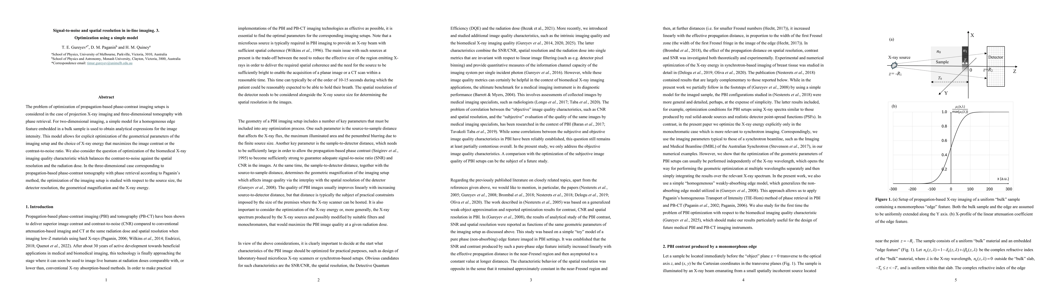
The problem of optimization of propagation-based phase-contrast imaging setups is considered in the case of projection X-ray imaging and three-dimensional tomography with phase retrieval. For two-dimensional imaging, a simple model for a homogeneous edge feature embedded in a bulk sample is used to obtain analytical expressions for the image intensity. This model allows for explicit optimization of the geometrical parameters of the imaging setup and the choice of X-ray energy that maximizes the image contrast or the contrast-to-noise ratio. We also consider the question of optimization of the biomedical X-ray imaging quality characteristic which balances the contrast-to-noise against the spatial resolution and the radiation dose. In the three-dimensional case corresponding to propagation-based phase-contrast tomography with phase retrieval according to Paganin's method, the optimization of the imaging setup is studied with respect to the source size, the detector resolution, the geometrical magnification and the X-ray energy.
AI Key Findings
Generated Oct 04, 2025
Methodology — What approach did the authors take?
The research employed analytical modeling and numerical simulations to derive expressions for contrast and spatial resolution in propagation-based phase-contrast imaging. It combined geometric optics principles with X-ray interaction physics to optimize imaging parameters.
Key Results — What are the main findings?
- Optimal X-ray wavelengths for maximizing biomedical image quality correspond to ~10% photon transmission through the sample
- Optimal magnification is determined by the ratio of X-ray source size to detector resolution
- Contrast, SNR, and CNR increase linearly with effective propagation distance in the near-Fresnel region
Significance — Why does this research matter?
This work provides actionable guidelines for optimizing X-ray imaging systems for biomedical applications, balancing image quality with radiation dose and system performance.
Technical Contribution — What is the technical contribution?
Derived explicit mathematical relationships between imaging quality metrics and system parameters, enabling analytical optimization of imaging configurations
Novelty — What is new about this work?
First comprehensive analytical framework that simultaneously optimizes spatial resolution, contrast, and biomedical image quality metrics while considering radiation dose and photon statistics
Limitations — What are the limitations of this study?
- Approximations in X-ray material interaction dependencies may limit broad applicability
- Results are based on monochromatic X-ray assumptions
- Optimization for polychromatic spectra requires additional considerations
Future Work — What did the authors propose for future work?
- Develop optimization frameworks for polychromatic X-ray spectra
- Validate results with experimental imaging systems
- Extend analysis to include detector quantum efficiency effects
- Investigate time-resolved phase-contrast imaging optimization
Paper Details
How to Cite This Paper
@article{paganin2025signal,
title = {Signal-to-noise and spatial resolution in in-line imaging. 3.
Optimization using a simple model},
author = {Paganin, D. M. and Quiney, H. M. and Gureyev, T. E.},
year = {2025},
eprint = {2510.01892},
archivePrefix = {arXiv},
primaryClass = {physics.med-ph},
}
Paganin, D., Quiney, H., & Gureyev, T. (2025). Signal-to-noise and spatial resolution in in-line imaging. 3.
Optimization using a simple model. arXiv. https://arxiv.org/abs/2510.01892
Paganin, D. M., et al. "Signal-to-noise and spatial resolution in in-line imaging. 3.
Optimization using a simple model." arXiv, 2025, arxiv.org/abs/2510.01892.
PDF Preview
Similar Papers
Found 4 papersSignal-to-noise and spatial resolution in in-line imaging. 2. Phase-contrast tomography
T. E. Gureyev, D. M. Paganin, K. M. Pavlov et al.
Signal-to-noise and spatial resolution in in-line imaging. 1. Basic theory, numerical simulations and planar experimental images
T. E. Gureyev, D. M. Paganin, H. M. Quiney
On spatial resolution, signal-to-noise and information capacity of linear imaging systems
Spatial resolution, noise and information in the computational-imaging era
David M. Paganin, Timur E. Gureyev, Alexander Kozlov
Comments (0)