Publication
Metrics
AI Quick Summary
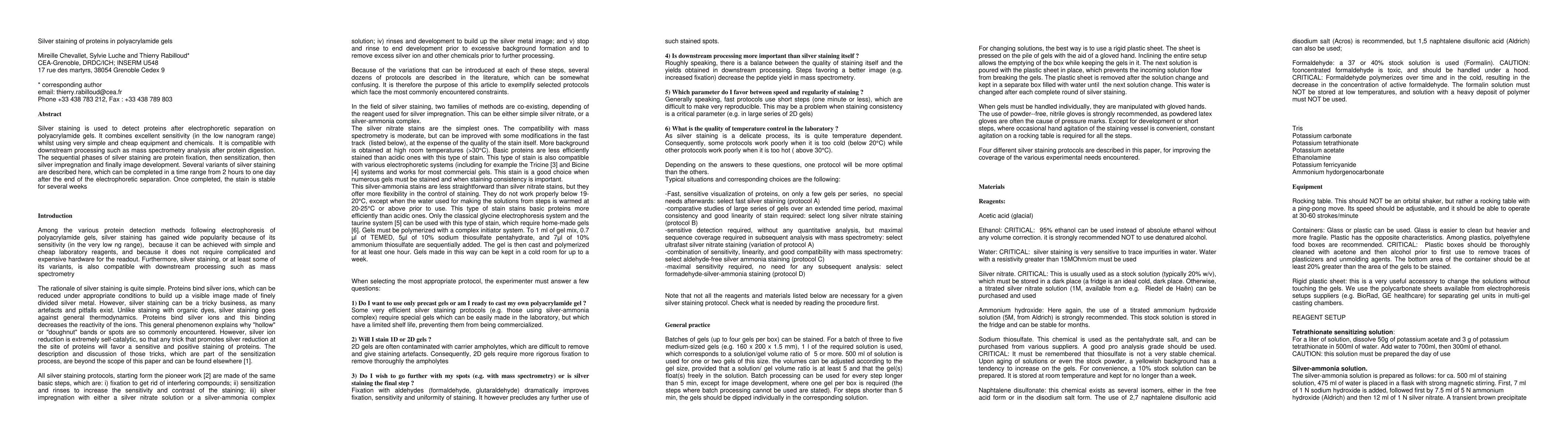
This paper describes a highly sensitive and cost-effective silver staining method for detecting proteins in polyacrylamide gels, compatible with downstream analyses like mass spectrometry. The process involves protein fixation, sensitization, silver impregnation, and image development, with variants allowing completion in 2 hours to 1 day.
Paper Preview
Abstract
Silver staining is used to detect proteins after electrophoretic separation on polyacrylamide gels. It combines excellent sensitivity (in the low nanogram range) with the use of very simple and cheap equipment and chemicals. It is compatible with downstream processing, such as mass spectrometry analysis after protein digestion. The sequential phases of silver staining are protein fixation, then sensitization, then silver impregnation and finally image development. Several variants of silver staining are described here, which can be completed in a time range from 2 h to 1 d after the end of the electrophoretic separation. Once completed, the stain is stable for several weeks.
AI Key Findings
Get AI-generated insights about this paper's methodology, results, significance, and more — seven facets brought into focus.
Impact
Paper Details
PDF Preview
Key Terms
Citation Network
Current paper (gray), citations (green), references (blue)
Display is limited for performance on very large graphs.
Discussion 0