Simplifying recombinant protein production: Combining Golden Gate cloning with a standardized protein purification scheme
Publication
Metrics
AI Quick Summary
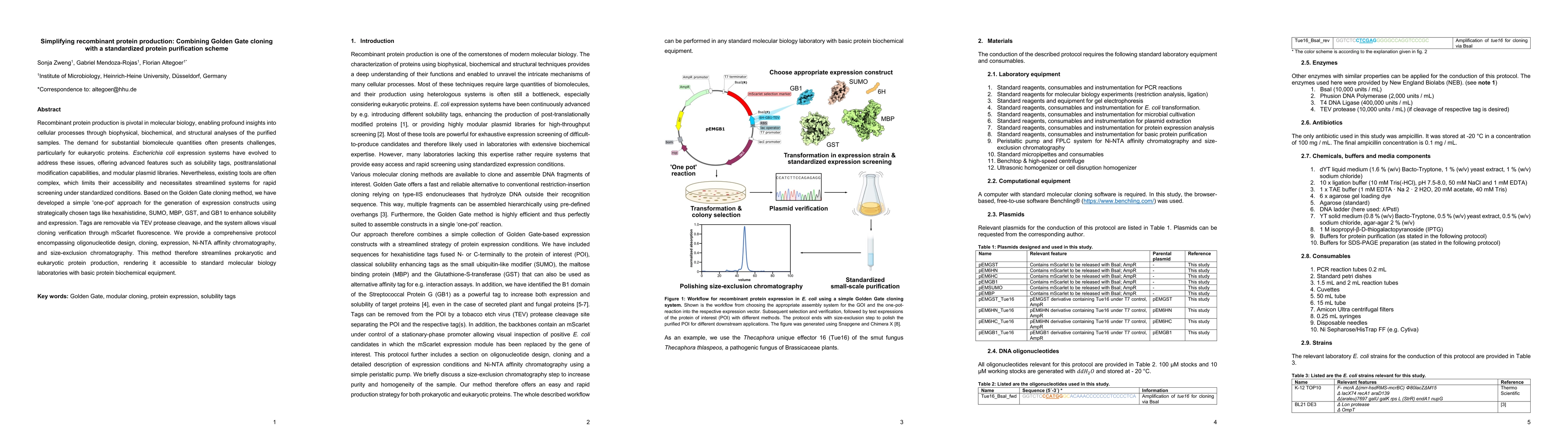
This paper presents a streamlined "one-pot" approach combining Golden Gate cloning with standardized protein purification to simplify recombinant protein production, featuring removable tags and visual cloning verification, thus making the process accessible to laboratories with basic equipment. The protocol includes detailed steps for expression, Ni-NTA chromatography, and size-exclusion chromatography.
Paper Preview
Abstract
Recombinant protein production is pivotal in molecular biology, enabling profound insights into cellular processes through biophysical, biochemical, and structural analyses of the purified samples. The demand for substantial biomolecule quantities often presents challenges, particularly for eukaryotic proteins. Escherichia coli expression systems have evolved to address these issues, offering advanced features such as solubility tags, posttranslational modification capabilities, and modular plasmid libraries. Nevertheless, existing tools are often complex, which limits their accessibility and necessitates streamlined systems for rapid screening under standardized conditions. Based on the Golden Gate cloning method, we have developed a simple 'one-pot' approach for the generation of expression constructs using strategically chosen tags like hexahistidine, SUMO, MBP, GST, and GB1 to enhance solubility and expression. Tags are removable via TEV protease cleavage, and the system allows visual cloning verification through mScarlet fluorescence. We provide a comprehensive protocol encompassing oligonucleotide design, cloning, expression, Ni-NTA affinity chromatography, and size-exclusion chromatography. This method therefore streamlines prokaryotic and eukaryotic protein production, rendering it accessible to standard molecular biology laboratories with basic protein biochemical equipment.
AI Key Findings
Get AI-generated insights about this paper's methodology, results, significance, and more — seven facets brought into focus.
Impact
Paper Details
Authors
PDF Preview
Key Terms
Citation Network
Current paper (gray), citations (green), references (blue)
Display is limited for performance on very large graphs.
Discussion 0