Simulation of an imaging system for internal contamination of lungs using MPA-MURA coded aperture collimator
Publication
Metrics
AI Quick Summary
This study employs the Monte Carlo method to simulate lung contamination imaging using an MPA-MURA coded-aperture collimator, achieving higher detection efficiency and a signal-to-noise ratio of 3.98 dB, despite lower spatial resolution compared to parallel-hole imaging.
Paper Preview
Abstract
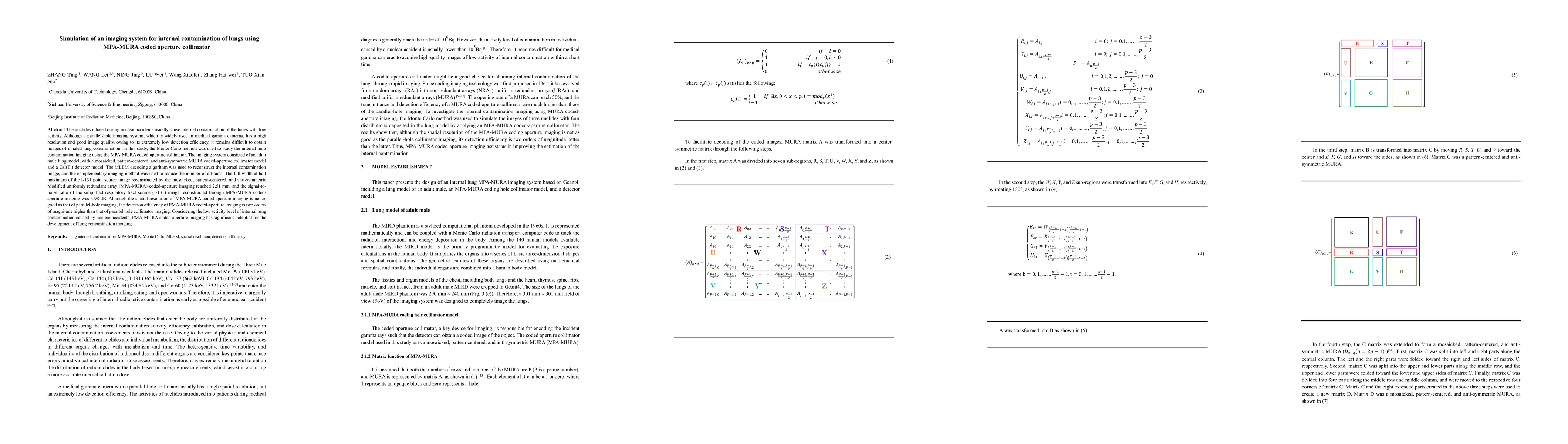
The nuclides inhaled during nuclear accidents usually cause internal contamination of the lungs with low activity. Although a parallel-hole imaging system, which is widely used in medical gamma cameras, has a high resolution and good image quality, owing to its extremely low detection efficiency, it remains difficult to obtain images of inhaled lung contamination. In this study, the Monte Carlo method was used to study the internal lung contamination imaging using the MPA-MURA coded-aperture collimator. The imaging system consisted of an adult male lung model, with a mosaicked, pattern-centered, and anti-symmetric MURA coded-aperture collimator model and a CsI(Tl) detector model. The MLEM decoding algorithm was used to reconstruct the internal contamination image, and the complementary imaging method was used to reduce the number of artifacts. The full width at half maximum of the I-131 point source image reconstructed by the mosaicked, pattern-centered, and anti-symmetric Modified uniformly redundant array (MPA-MURA) coded-aperture imaging reached 2.51 mm, and the signal-to-noise ratio of the simplified respiratory tract source (I-131) image reconstructed through MPA-MURA coded-aperture imaging was 3.98 dB. Although the spatial resolution of MPA-MURA coded aperture imaging is not as good as that of parallel-hole imaging, the detection efficiency of PMA-MURA coded-aperture imaging is two orders of magnitude higher than that of parallel hole collimator imaging. Considering the low activity level of internal lung contamination caused by nuclear accidents, PMA-MURA coded-aperture imaging has significant potential for the development of lung contamination imaging.
AI Key Findings
Get AI-generated insights about this paper's methodology, results, significance, and more — seven facets brought into focus.
Impact
Paper Details
Authors
PDF Preview
Key Terms
Citation Network
Current paper (gray), citations (green), references (blue)
Display is limited for performance on very large graphs.
Discussion 0