Quick Summary:

Researchers developed an algorithm to simulate brain resection cavities for image segmentation, achieving comparable accuracy to human annotations without manual data labeling, promising a new approach for improving resective surgery outcomes.

Summary

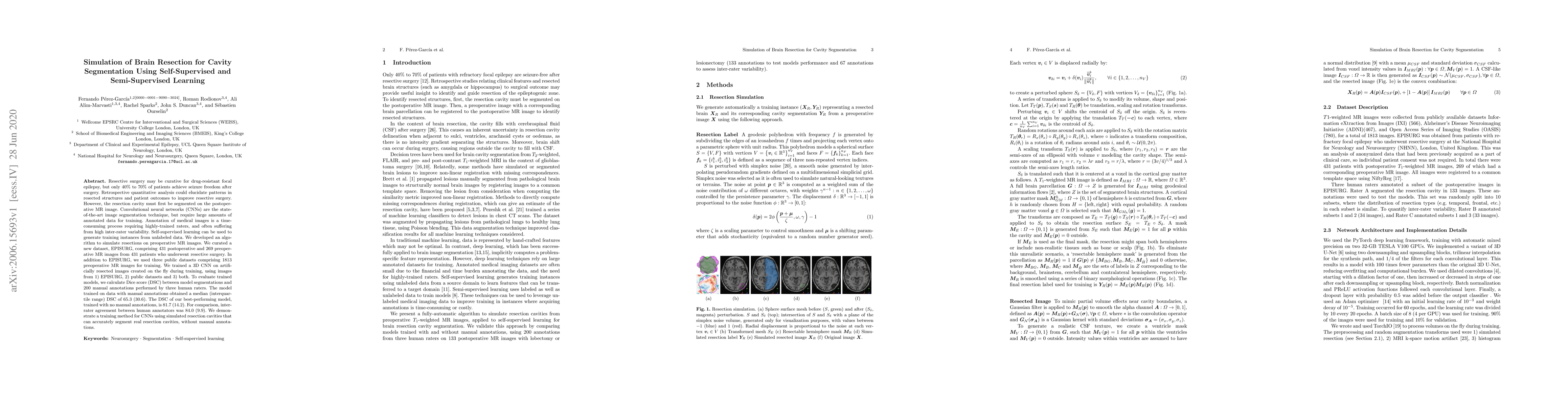
Resective surgery may be curative for drug-resistant focal epilepsy, but only 40% to 70% of patients achieve seizure freedom after surgery. Retrospective quantitative analysis could elucidate patterns in resected structures and patient outcomes to improve resective surgery. However, the resection cavity must first be segmented on the postoperative MR image. Convolutional neural networks (CNNs) are the state-of-the-art image segmentation technique, but require large amounts of annotated data for training. Annotation of medical images is a time-consuming process requiring highly-trained raters, and often suffering from high inter-rater variability. Self-supervised learning can be used to generate training instances from unlabeled data. We developed an algorithm to simulate resections on preoperative MR images. We curated a new dataset, EPISURG, comprising 431 postoperative and 269 preoperative MR images from 431 patients who underwent resective surgery. In addition to EPISURG, we used three public datasets comprising 1813 preoperative MR images for training. We trained a 3D CNN on artificially resected images created on the fly during training, using images from 1) EPISURG, 2) public datasets and 3) both. To evaluate trained models, we calculate Dice score (DSC) between model segmentations and 200 manual annotations performed by three human raters. The model trained on data with manual annotations obtained a median (interquartile range) DSC of 65.3 (30.6). The DSC of our best-performing model, trained with no manual annotations, is 81.7 (14.2). For comparison, inter-rater agreement between human annotators was 84.0 (9.9). We demonstrate a training method for CNNs using simulated resection cavities that can accurately segment real resection cavities, without manual annotations.

AI Key Findings

Get AI-generated insights about this paper's methodology, results, and significance.

Paper Details

PDF Preview

Download PDF

Key Terms

annotations (0.281)
manual (0.265)
images (0.211)
trained (0.208)
cavities (0.201)
cnns (0.199)
training (0.186)
patients (0.183)
inter (0.160)
public (0.157)
human (0.139)
image (0.125)
datasets (0.122)
drug (0.118)
elucidate (0.118)
patient (0.115)
amounts (0.113)
annotated (0.111)
self supervised (0.110)
median (0.110)

Citation Network

Current paper (gray), citations (green), references (blue)

Current Paper
Citations
References
Click to view
Title Authors Year Actions
BrainMorph: A Foundational Keypoint Model for Robust and Flexible Brain MRI Registration
Alan Q. Wang, Rachit Saluja, Heejong Kim, Xinzi He, Adrian V. Dalca, M. Sabuncu
2025
View Paper
BrainMorph: A Foundational Keypoint Model for Robust and Flexible Brain MRI Registration
Alan Q. Wang, Rachit Saluja, Heejong Kim, Xinzi He, Adrian V. Dalca, M. Sabuncu
2024
View Paper
Matpr-Unet: A Multi Attention Two-Path Residual Unet for Focal Cortical Dysplasia Lesions Segmentation
Wenjing Zhang, Hao Yu, Manli Zhang, Gongpeng Cao, Guixia Kang, Lixin Cai
2024
View Paper
Fully automatic segmentation of brain lacunas resulting from resective surgery using a 3D deep learning model
R. Casseb, B. D. de Campos, Wallace Souza Loos, Marcelo Eduardo Ramos Barbosa, M. Alvim, Gabriel Chagas Lutfala Paulino, Francesco Pucci, Samuel Worrell, Roberto Medeiros de Souza, Lara Jehi, F. Cendes
2023
View Paper
Multi-layer Aggregation as a key to feature-based OOD detection
Benjamin Lambert, Florence Forbes, Senan Doyle, M. Dojat
2023
View Paper
Deep learning-based automated segmentation of resection cavities on postsurgical epilepsy MRI
T. Arnold, Ramya Muthukrishnan, A. Pattnaik, N. Sinha, Adam D. Gibson, Hannah Gonzalez, Sandhitsu R. Das, B. Litt, Dario J. Englot, V. Morgan, K. Davis, J. Stein
2022
View Paper
Epidemic models characterize seizure propagation and the effects of epilepsy surgery in individualized brain networks based on MEG and invasive EEG recordings
Ana P Millán, Elisabeth C. W. van Straaten, C. Stam, I. A. Nissen, S. Idema, J. C. Baayen, P. van Mieghem, A. Hillebrand
2021
View Paper
A self-supervised learning strategy for postoperative brain cavity segmentation simulating resections
Fernando Pérez-García, R. Dorent, M. Rizzi, F. Cardinale, Valerio Frazzini, Vincent Navarro, Caroline Essert, I. Ollivier, T. Vercauteren, R. Sparks, J. Duncan, S. Ourselin
2021
View Paper
A generative model of hyperelastic strain energy density functions for multiple tissue brain deformation
Alejandro Granados, Fernando Pérez-García, M. Schweiger, V. Vakharia, S. Vos, A. Miserocchi, A. McEvoy, J. Duncan, R. Sparks, S. Ourselin
2020
View Paper
TorchIO: A Python library for efficient loading, preprocessing, augmentation and patch-based sampling of medical images in deep learning
Fernando Pérez-García, R. Sparks, S. Ourselin
2020
View Paper
Computer Methods and Programs in Biomedicine
J. E. Herrera-Serrano, J. Macías-Díaz, I. Medina-Ramírez, J. A. Guerrero
View Paper

Comments (0)

This website uses cookies to ensure you get the best experience. Learn more