Summary
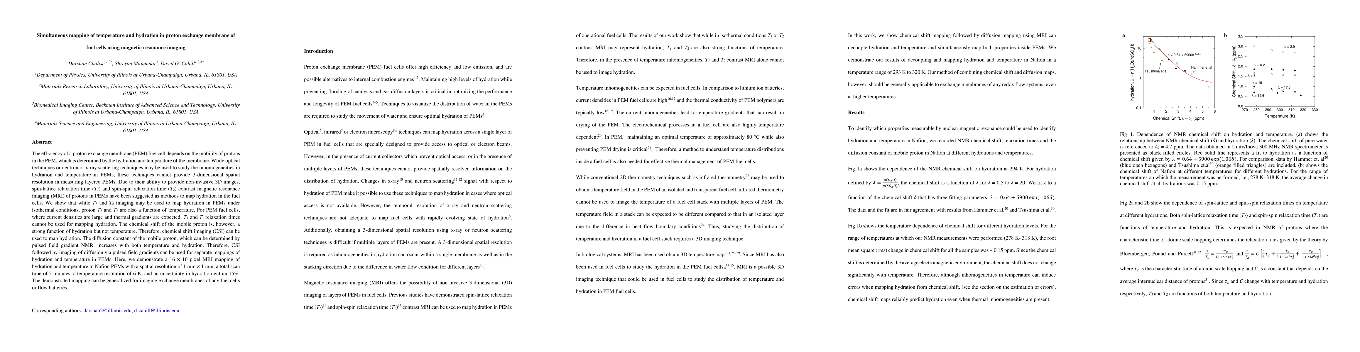
The efficiency of a proton exchange membrane (PEM) fuel cell depends on the mobility of protons in the PEM, which is determined by the hydration and temperature of the membrane. While optical techniques or neutron or x-ray scattering techniques may be used to study the inhomogeneities in hydration and temperature in PEMs, these techniques cannot provide 3 dimensional spatial resolution in measuring layered PEMs. Due to their ability to provide non-invasive 3D images, spin-lattice relaxation time (T1) and spin-spin relaxation time (T2) contrast magnetic resonance imaging (MRI) of protons in PEMs have been suggested as methods to map hydration in the fuel cells. We show that while T1 and T2 imaging may be used to map hydration in PEMs under isothermal conditions, proton T1 and T2 are also a function of temperature. For PEM fuel cells, where current densities are large and thermal gradients are expected, T1 and T2 relaxation times cannot be used for mapping hydration. The chemical shift of the mobile proton is, however, a strong function of hydration but not temperature. Therefore, chemical shift imaging (CSI) can be used to map hydration. The diffusion constant of the mobile proton, which can be determined by pulsed field gradient NMR, increases with both temperature and hydration. Therefore, CSI followed by imaging of diffusion via pulsed field gradients can be used for separate mappings of hydration and temperature in PEMs. Here, we demonstrate a 16 x 16 pixel MRI mapping of hydration and temperature in Nafion PEMs with a spatial resolution of 1 mm x 1 mm, a total scan time of 3 minutes, a temperature resolution of 6 K, and an uncertainty in hydration within 15%. The demonstrated mapping can be generalized for imaging exchange membranes of any fuel cells or flow batteries.
AI Key Findings
Generated Sep 03, 2025
Methodology
The research utilizes magnetic resonance imaging (MRI) to simultaneously map hydration and temperature in proton exchange membrane fuel cells (PEMFCs), employing chemical shift imaging (CSI) for hydration and pulsed field gradient NMR for diffusion constant mapping, followed by temperature reconstruction based on the diffusion constant and expected values at a reference temperature.
Key Results
- Simultaneous 16 x 16 pixel mapping of hydration and temperature in Nafion PEMFCs with 1 mm x 1 mm spatial resolution achieved in 3 minutes.
- Temperature resolution of 6 K and hydration uncertainty within 15%.
- Demonstration of the method's applicability to other fuel cell or flow battery exchange membranes.
Significance
This research provides a non-invasive technique to understand and optimize PEMFC performance by mapping hydration and temperature inhomogeneities, which are crucial for proton mobility and efficiency.
Technical Contribution
Development of an MRI technique to decouple and map both temperature and hydration in PEMFCs, applicable to various redox flow systems.
Novelty
The method overcomes limitations of previous techniques by providing 3D spatial resolution for measuring layered PEMFCs, using CSI for hydration and diffusion mapping for temperature, enabling simultaneous and accurate mapping of critical parameters for PEMFC performance.
Limitations
- Current system's noise contributing to uncertainties in hydration and temperature measurements.
- Longer scan times for larger fields of view or higher spatial resolutions.
Future Work
- Optimization of MRI system and data acquisition for improved resolution and reduced uncertainties.
- Application of faster spectroscopic imaging techniques, like echo-planar imaging, to reduce scan times for larger or more complex fuel cell configurations.
Paper Details
PDF Preview
Key Terms
Citation Network
Current paper (gray), citations (green), references (blue)
Display is limited for performance on very large graphs.
Similar Papers
Found 4 papersNMR contributions to the study of water transfer in proton exchange membranes for fuel cells
Jean-Christophe Perrin, Assma El Kaddouri, Laouès Guendouz et al.
A Critical Review of Proton Exchange Membrane Fuel Cells Matter Transports and Voltage Polarisation for Modelling
Raphaël Gass, Zhongliang Li, Rachid Outbib et al.
| Title | Authors | Year | Actions |
|---|
Comments (0)