Publication

Published: Sep 19, 2025
Categories: physics.med-ph, physics.app-ph, physics.optics

Metrics

Source: ArXiv

Quick Actions

View PDF

Quick Answers

What methodology did the authors use?

The study developed a multispectral imaging system integrating synchronized color cameras with custom filters and a beam splitter to capture white light reflectance and multiple fluorescence signals in real-time. It used phantoms simulating clinical conditions to evaluate performance, employing linear unmixing algorithms for fluorescence separation and Delta E metrics for color fidelity assessment. More in Methodology →

What are the key results?

The system successfully separated fluorescence signals from PpIX, fluorescein sodium, and ICG in real-time with minimal crosstalk. — Reflectance images showed high color fidelity with Delta E values < 2 for most color patches, indicating imperceptible to barely perceptible differences compared to conventional cameras. More in Key Results →

Why is this work significant?

This research addresses fragmented intraoperative imaging by providing real-time, integrated visualization of anatomical and fluorescence data, improving surgical precision, reducing cognitive load, and supporting more efficient and accurate interventions. More in Significance →

What are the main limitations?

The system was tested using phantoms rather than real clinical scenarios, limiting direct applicability to actual surgical environments. — Further studies are needed to validate performance under conditions like tissue autofluorescence and varying optical properties. More in Limitations →

Paper Preview

Abstract

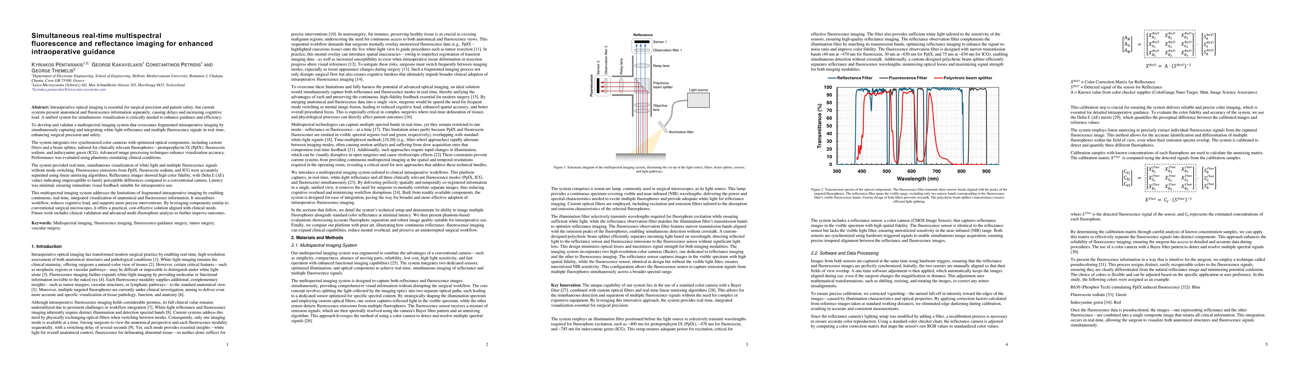
Intraoperative optical imaging is essential for surgical precision and patient safety, but current systems present anatomical and fluorescence information separately, causing delays and increasing cognitive load. A unified system for simultaneous visualization is critically needed to enhance guidance and efficiency. To develop and validate a multispectral imaging system that overcomes fragmented intraoperative imaging by simultaneously capturing and integrating white light reflectance and multiple fluorescence signals in real-time, enhancing surgical precision and safety. The system integrates two synchronized color cameras with optimized optical components, including custom filters and a beam splitter, tailored for clinically relevant fluorophores protoporphyrin IX (PpIX), fluorescein sodium, and indocyanine green (ICG). Advanced image processing techniques enhance visualization accuracy. Performance was evaluated using phantoms simulating clinical conditions. The system provided real time, simultaneous visualization of white light and multiple fluorescence signals without mode switching. Fluorescence emissions from PpIX, fluorescein sodium, and ICG were accurately separated using linear unmixing algorithms. Reflectance images showed high color fidelity, with Delta E values indicating imperceptible to barely perceptible differences compared to a conventional camera. Latency was minimal, ensuring immediate visual feedback suitable for intraoperative use. This multispectral imaging system addresses the limitations of fragmented intraoperative imaging by enabling continuous, real time, integrated visualization of anatomical and fluorescence information. It streamlines workflow, reduces cognitive load, and supports more precise interventions. By leveraging components similar to conventional surgical microscopes, it offers a practical, cost effective solution aligned with clinical needs.

AI Key Findings

Generated Sep 30, 2025

Methodology — What approach did the authors take?

The study developed a multispectral imaging system integrating synchronized color cameras with custom filters and a beam splitter to capture white light reflectance and multiple fluorescence signals in real-time. It used phantoms simulating clinical conditions to evaluate performance, employing linear unmixing algorithms for fluorescence separation and Delta E metrics for color fidelity assessment.

Key Results — What are the main findings?

  • The system successfully separated fluorescence signals from PpIX, fluorescein sodium, and ICG in real-time with minimal crosstalk.
  • Reflectance images showed high color fidelity with Delta E values < 2 for most color patches, indicating imperceptible to barely perceptible differences compared to conventional cameras.
  • Composite imaging combined reflectance and fluorescence data into a single view, enabling simultaneous visualization of anatomical structures and fluorescence information.

Significance — Why does this research matter?

This research addresses fragmented intraoperative imaging by providing real-time, integrated visualization of anatomical and fluorescence data, improving surgical precision, reducing cognitive load, and supporting more efficient and accurate interventions.

Technical Contribution — What is the technical contribution?

The system integrates hardware components (color cameras, filters, beam splitter) with advanced image processing algorithms to achieve real-time multispectral fluorescence and reflectance imaging, enabling seamless integration of multiple imaging modalities.

Novelty — What is new about this work?

The system provides simultaneous, real-time visualization of white light reflectance and multiple fluorescence signals without mode switching, overcoming limitations of time-multiplexed systems and offering a practical, cost-effective solution for intraoperative guidance.

Limitations — What are the limitations of this study?

  • The system was tested using phantoms rather than real clinical scenarios, limiting direct applicability to actual surgical environments.
  • Further studies are needed to validate performance under conditions like tissue autofluorescence and varying optical properties.

Future Work — What did the authors propose for future work?

  • Conduct in vivo clinical trials to assess system performance in real surgical settings.
  • Expand spectral range and compatibility to accommodate emerging fluorophores.
  • Investigate compact and portable designs for broader accessibility in resource-limited settings.

Paper Details

Paper ID: 2509.15979
Comments: 15 pages, 7 Figures
Categories:
physics.med-ph physics.app-ph physics.optics

How to Cite This Paper

BibTeX
@article{petridis2025simultaneous,
  title         = {Simultaneous real-time multispectral fluorescence and reflectance
  imaging for enhanced intraoperative guidance},
  author        = {Petridis, Constantinos and Kakavelakis, George and Themelis, George and others},
  year          = {2025},
  eprint        = {2509.15979},
  archivePrefix = {arXiv},
  primaryClass  = {physics.med-ph},
}
APA 7
Petridis, C., Kakavelakis, G., Themelis, G., & Pentarakis, K. (2025). Simultaneous real-time multispectral fluorescence and reflectance
  imaging for enhanced intraoperative guidance. arXiv. https://arxiv.org/abs/2509.15979
MLA 9
Petridis, Constantinos, et al. "Simultaneous real-time multispectral fluorescence and reflectance
  imaging for enhanced intraoperative guidance." arXiv, 2025, arxiv.org/abs/2509.15979.

Comments (0)

This website uses cookies to ensure you get the best experience. Learn more