Publication
Metrics
AI Quick Summary
This paper introduces a single-pixel coherent diffraction imaging technique that extends the working spectrum to a wide waveband by using a single-pixel detector instead of a dense array, significantly reducing measurement dynamic range. The method successfully recovers 2D amplitude and phase maps from 1D intensity measurements without prior object information, validated with both phase objects and biological samples.
Paper Preview
Abstract
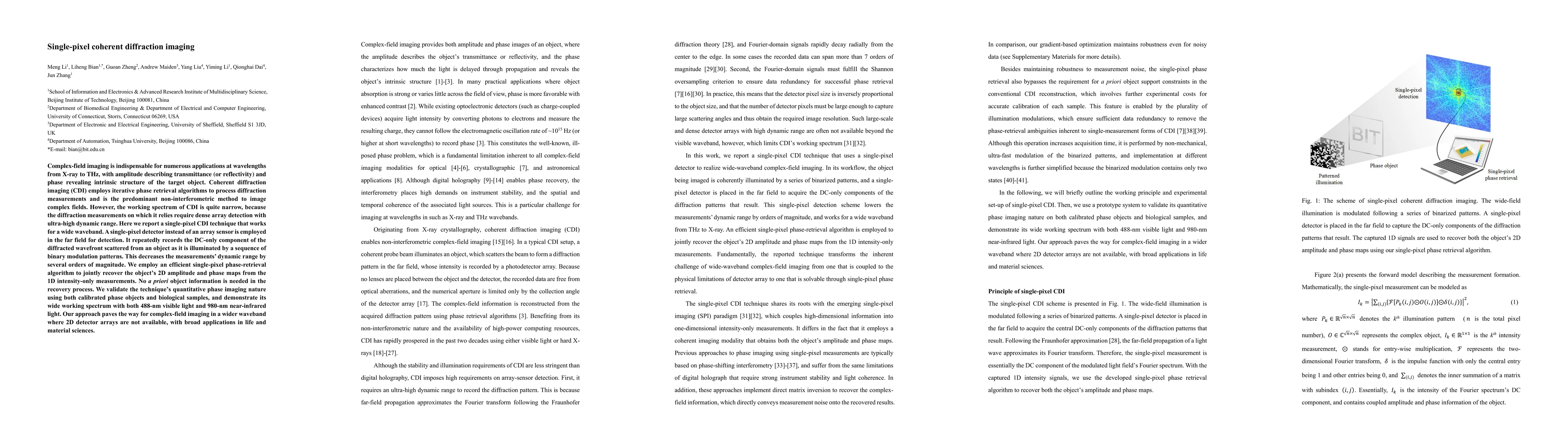
Complex-field imaging is indispensable for numerous applications at wavelengths from X-ray to THz, with amplitude describing transmittance (or reflectivity) and phase revealing intrinsic structure of the target object. Coherent diffraction imaging (CDI) employs iterative phase retrieval algorithms to process diffraction measurements and is the predominant non-interferometric method to image complex fields. However, the working spectrum of CDI is quite narrow, because the diffraction measurements on which it relies require dense array detection with ultra-high dynamic range. Here we report a single-pixel CDI technique that works for a wide waveband. A single-pixel detector instead of an array sensor is employed in the far field for detection. It repeatedly records the DC-only component of the diffracted wavefront scattered from an object as it is illuminated by a sequence of binary modulation patterns. This decreases the measurements' dynamic range by several orders of magnitude. We employ an efficient single-pixel phase-retrieval algorithm to jointly recover the object's 2D amplitude and phase maps from the 1D intensity-only measurements. No a priori object information is needed in the recovery process. We validate the technique's quantitative phase imaging nature using both calibrated phase objects and biological samples, and demonstrate its wide working spectrum with both 488-nm visible light and 980-nm near-infrared light. Our approach paves the way for complex-field imaging in a wider waveband where 2D detector arrays are not available, with broad applications in life and material sciences.
AI Key Findings
Get AI-generated insights about this paper's methodology, results, significance, and more — seven facets brought into focus.
Impact
Paper Details
Authors
PDF Preview
Key Terms
Citation Network
Current paper (gray), citations (green), references (blue)
Display is limited for performance on very large graphs.
Discussion 0