Single-Shot 3D Diffractive Imaging of Core-Shell Nanoparticles with Elemental Specificity
Publication
Metrics
AI Quick Summary
This paper presents a method for 3D coherent diffractive imaging of Au/Pd core-shell nanoparticles using femtosecond x-ray pulses, achieving 6 nm resolution. The technique successfully reconstructs 3D electron density from single-shot diffraction patterns, determining core and shell dimensions with high accuracy.
Paper Preview
Abstract
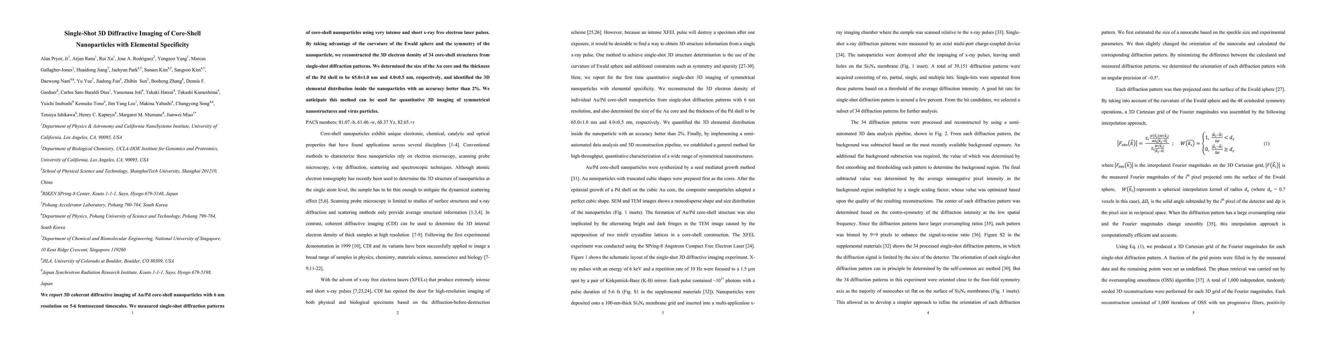
We report 3D coherent diffractive imaging of Au/Pd core-shell nanoparticles with 6 nm resolution on 5-6 femtosecond timescales. We measured single-shot diffraction patterns of core-shell nanoparticles using very intense and short x-ray free electron laser pulses. By taking advantage of the curvature of the Ewald sphere and the symmetry of the nanoparticle, we reconstructed the 3D electron density of 34 core-shell structures from single-shot diffraction patterns. We determined the size of the Au core and the thickness of the Pd shell to be 65.0 +/- 1.0 nm and 4.0 +/- 0.5 nm, respectively, and identified the 3D elemental distribution inside the nanoparticles with an accuracy better than 2%. We anticipate this method can be used for quantitative 3D imaging of symmetrical nanostructures and virus particles.
AI Key Findings
Get AI-generated insights about this paper's methodology, results, significance, and more — seven facets brought into focus.
Impact
Paper Details
PDF Preview
Key Terms
Citation Network
Current paper (gray), citations (green), references (blue)
Display is limited for performance on very large graphs.
Discussion 0