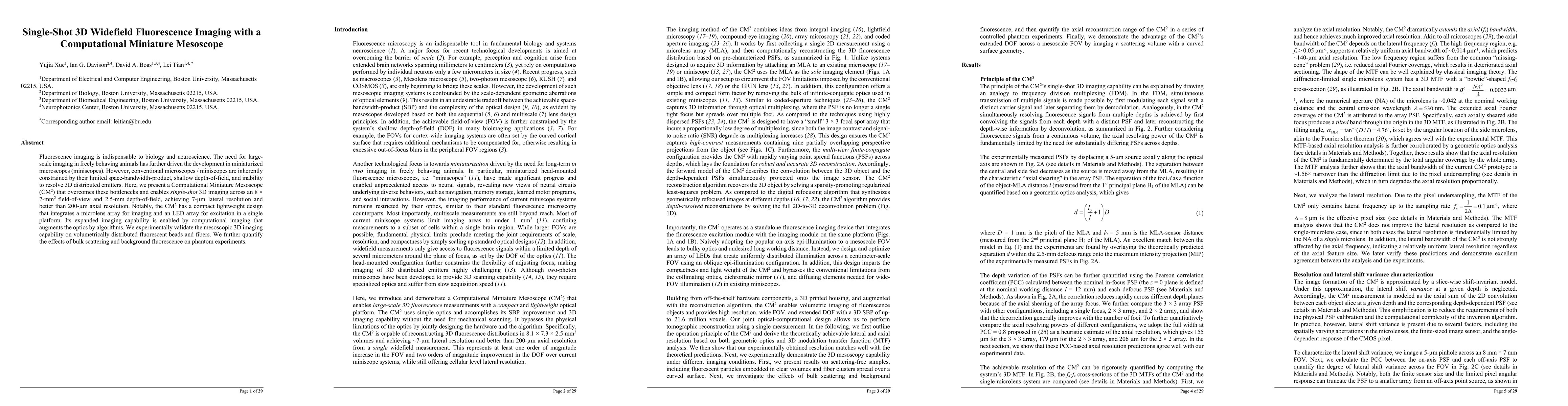
Single-Shot 3D Widefield Fluorescence Imaging with a Computational Miniature Mesoscope
Publication
Metrics
AI Quick Summary
This paper introduces a Computational Miniature Mesoscope (CM$^2$) that enables single-shot 3D widefield fluorescence imaging with high spatial and depth resolution, overcoming limitations of conventional miniscopes. The CM$^2$ integrates microlens and LED arrays in a compact design, validated through experiments on fluorescent beads and fibers.
Paper Preview
Abstract
Fluorescence imaging is indispensable to biology and neuroscience. The need for large-scale imaging in freely behaving animals has further driven the development in miniaturized microscopes (miniscopes). However, conventional microscopes / miniscopes are inherently constrained by their limited space-bandwidth-product, shallow depth-of-field, and the inability to resolve 3D distributed emitters. Here, we present a Computational Miniature Mesoscope (CM$^2$) that overcomes these bottlenecks and enables single-shot 3D imaging across an 8 $\times$ 7-mm$^2$ field-of-view and 2.5-mm depth-of-field, achieving 7-$\mu$m lateral resolution and better than 200-$\mu$m axial resolution. Notably, the CM$^2$ has a compact lightweight design that integrates a microlens array for imaging and an LED array for excitation in a single platform. Its expanded imaging capability is enabled by computational imaging that augments the optics by algorithms. We experimentally validate the mesoscopic 3D imaging capability on volumetrically distributed fluorescent beads and fibers. We further quantify the effects of bulk scattering and background fluorescence on phantom experiments.
AI Key Findings
Get AI-generated insights about this paper's methodology, results, significance, and more — seven facets brought into focus.
Impact
Paper Details
PDF Preview
Key Terms
Citation Network
Current paper (gray), citations (green), references (blue)
Display is limited for performance on very large graphs.
Discussion 0