ArxivLens

Quick Summary:

This paper introduces polarized shearing interference microscopy (PSIM), a single-shot quantitative imaging technique for capturing the dynamics of molecular orientation in birefringent materials at millisecond timescales. The method demonstrates high accuracy and speed, imaging a flowing liquid crystal at 506 frames per second, and holds promise for dynamic material characterization.

arXiv Id: 2106.06118
Date Published: 2021-06-14
Date Updated: 2021-06-14

Key Terms (TF-IDF):

orientation (0.259)
imaging (0.245)
polarization (0.225)
quantitative (0.220)
microscopy (0.210)
mu (0.180)
molecular (0.169)
light (0.141)
optically (0.137)
time scale (0.135)
performances (0.134)
validated (0.133)
dynamics (0.131)
extracting (0.131)
frames (0.129)
diffraction (0.129)
steady state (0.127)
camera (0.123)
shot (0.123)
steady (0.121)
License: http://creativecommons.org/licenses/by/4.0/

Summary:

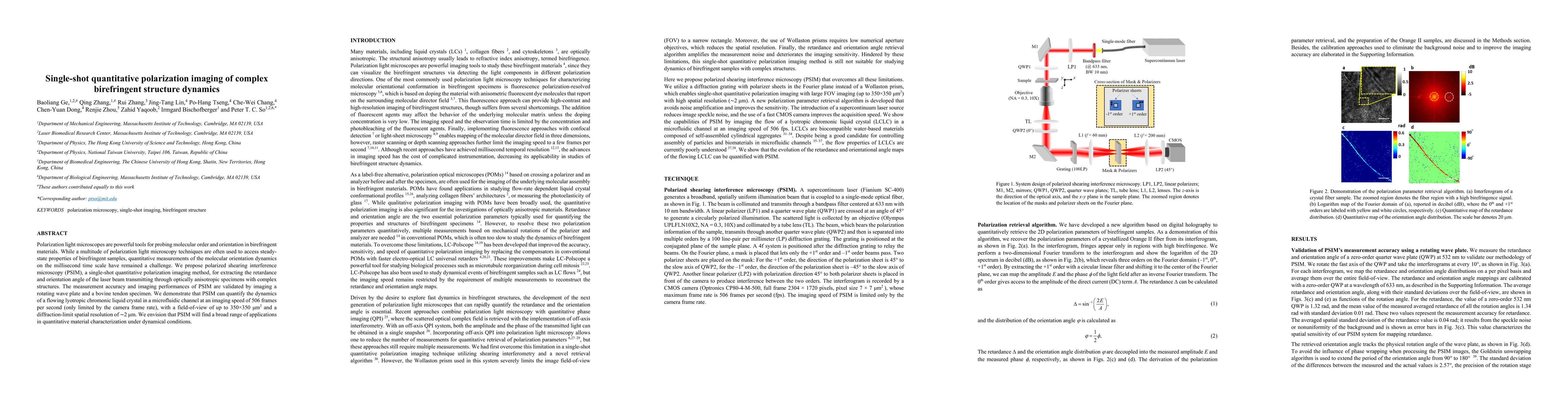
Polarization light microscopes are powerful tools for probing molecular order and orientation in birefringent materials. While a multitude of polarization light microscopy techniques are often used to access steady-state properties of birefringent samples, quantitative measurements of the molecular orientation dynamics on the millisecond time scale have remained a challenge. We propose polarized shearing interference microscopy (PSIM), a single-shot quantitative polarization imaging method, for extracting the retardance and orientation angle of the laser beam transmitting through optically anisotropic specimens with complex structures. The measurement accuracy and imaging performances of PSIM are validated by imaging a rotating wave plate and a bovine tendon specimen. We demonstrate that PSIM can quantify the dynamics of a flowing lyotropic chromonic liquid crystal in a microfluidic channel at an imaging speed of 506 frames per second (only limited by the camera frame rate), with a field-of-view of up to $350\times350 \mu m^2$ and a diffraction-limit spatial resolution of $\sim 2\mu m$. We envision that PSIM will find a broad range of applications in quantitative material characterization under dynamical conditions.

Citation & Reference Network Visualization

This graph shows how this paper is connected to other research. The current paper is shown in dark gray, papers that cite it are in green, and papers it references are in blue. You can:

Current Paper
Papers citing this work (Citations)
Papers cited by this work (References)

• Hover over any node to see the paper title

• Click a node to view that paper's details

• Drag nodes to rearrange the graph

• Use the zoom controls (bottom right) to zoom in/out

Citations and References

Title Authors Year Actions
Polarization digital holographic microscope for live cell imaging
Rakesh Kumar Singh, Shivam Chaubey, Mohit Rathor, Rupen Tamang, Biplob Koch
2024
View Paper
Real‐Time Jones Matrix Synthesis by Compact Polarization Inline Holographic Microscopy
Hanzi Liu, Vinu R.V., Kaiquan Chen, Dongyang Liao, Ziyang Chen, Jixiong Pu
2024
View Paper
Visualizing 3D Anisotropic Molecular Orientation in Polarization Holographic Optical Elements via Dielectric Tensor Tomography
Juheon Lee, Heeju Son, Seung Jae Hong, Hervé Hugonnet, Joona Bang, Seungwoo Lee, YongKeun Park
2024
View Paper
Flow-induced periodic chiral structures in an achiral nematic liquid crystal
Qing Zhang, Weiqiang Wang, Shuang Zhou, Rui Zhang, Irmgard Bischofberger
2024
View Paper
Single-Shot Quantitative Polarization Imaging of Live Cancer Cells
Mohit Rathor, Shivam Chaubey, Rupen Tamang, Biplob Koch, Rakesh Kumar Singh
2023
View Paper
Polarization-sensitive intensity diffraction tomography
Seungri Song, Jeongsoo Kim, T. Moon, Baekcheon Seong, Woojun Kim, Chang-Hyuk Yoo, Jun-Kyu Choi, C. Joo
2023
View Paper
Tensorial tomographic Fourier ptychography with applications to muscle tissue imaging
Shiqi Xu, Xiang Dai, P. Ritter, K. Lee, Xi Yang, L. Kreiss, Kevin C Zhou, Kanghyun Kim, A. Chaware, Jadee L. Neff, C. Glass, Seung Ah Lee, O. Friedrich, R. Horstmeyer
2023
View Paper
High-resolution assessment of multidimensional cellular mechanics using label-free refractive-index traction force microscopy
Moosung Lee, Hyuntae Jeong, Chaeyeon Lee, Mahn Jae Lee, Benedict Reve Delmo, Won Do Heo, J. Shin, Yongkeun Park
2023
View Paper
Dendritic patterns from shear-enhanced anisotropy in nematic liquid crystals
Qing Zhang, Shuang Zhou, Rui Zhang, Irmgard Bischofberger
2023
View Paper
Tensorial tomographic differential phase-contrast microscopy
Shiqi Xu, Xiang Dai, Xi Yang, Kevin C Zhou, Kanghyun Kim, Vinayak Pathak, C. Glass, R. Horstmeyer
2022
View Paper
Quantitative Jones matrix imaging using vectorial Fourier ptychography.
Xiang Dai, Shiqi Xu, Xi Yang, Kevin C Zhou, C. Glass, P. C. Konda, R. Horstmeyer
2021
View Paper
Structures and topological defects in pressure-driven lyotropic chromonic liquid crystals
Qing Zhang, Rui Zhang, Baoliang Ge, Z. Yaqoob, Peter T. C. So, Irmgard Bischofberger
2021
View Paper
Microsphere-assisted single-shot polarization holographic microscopy for quantitative birefringence imaging of dynamic samples
Qingyang Yue, Hao Ma, Ya-Bo Han, Hongyi Huang, Xianlong Liu, Yang Yang, Cheng-Shan Guo
2025
View Paper
高灵敏度干涉定量相位显微前沿进展与应用(封面文章·特邀)
周楠森 ZHOU Nansen, 吴沐蕾 WU Mulei, 聂宇洁 NIE Yujie, 程加雨 CHENG Jiayu, 周仁杰 ZHOU Renjie
2024
View Paper

Comments (0)

This website uses cookies to ensure you get the best experience. Learn more