Publication
Metrics
AI Quick Summary
This paper discusses the challenges of skull-stripping in tumor-bearing brain images, highlighting the inadequacies of existing algorithms. The authors propose a new method tailored for this specific task, addressing issues like anisotropic resolution and partial coverage in clinical MRIs.
Paper Preview
Abstract
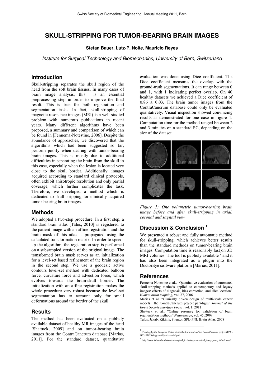
Skull-stripping separates the skull region of the head from the soft brain tissues. In many cases of brain image analysis, this is an essential preprocessing step in order to improve the final result. This is true for both registration and segmentation tasks. In fact, skull-stripping of magnetic resonance images (MRI) is a well-studied problem with numerous publications in recent years. Many different algorithms have been proposed, a summary and comparison of which can be found in [Fennema-Notestine, 2006]. Despite the abundance of approaches, we discovered that the algorithms which had been suggested so far, perform poorly when dealing with tumor-bearing brain images. This is mostly due to additional difficulties in separating the brain from the skull in this case, especially when the lesion is located very close to the skull border. Additionally, images acquired according to standard clinical protocols, often exhibit anisotropic resolution and only partial coverage, which further complicates the task. Therefore, we developed a method which is dedicated to skull-stripping for clinically acquired tumor-bearing brain images.
AI Key Findings
Get AI-generated insights about this paper's methodology, results, significance, and more — seven facets brought into focus.
Impact
Paper Details
PDF Preview
Key Terms
Citation Network
Current paper (gray), citations (green), references (blue)
Display is limited for performance on very large graphs.
Discussion 0