Space rocks and optimising scanning electron channelling contrast
Publication
Metrics
AI Quick Summary
The study confirmed the relationship between detector positioning and image contrast modes, providing new insights into forescatter electron imaging.
Paper Preview
Abstract
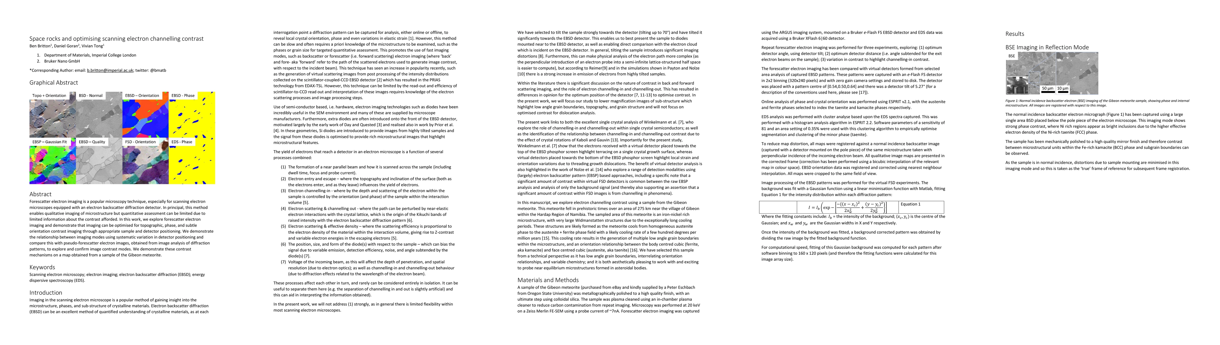
Forescatter electron imaging is a popular microscopy technique, especially for scanning electron microscopes equipped with an electron backscatter diffraction detector. In principal, this method enables qualitative imaging of microstructure but quantitative assessment can be limited due to limited information about the contrast afforded. In this work, we explore forescatter electron imaging and demonstrate that imaging can be optimised for topographic, phase, and subtle orientation contrast imaging through appropriate sample and detector positioning. We demonstrate the relationship between imaging modes using systematic variation in detector positioning and compare this with pseudo-forescatter electron images, obtained from image analysis of diffraction patterns, to explore and confirm image contrast modes. We demonstrate these contrast mechanisms on a map obtained from a sample of the Gibeon meteorite.
AI Key Findings
Get AI-generated insights about this paper's methodology, results, significance, and more — seven facets brought into focus.
Impact
Paper Details
PDF Preview
Key Terms
Citation Network
Current paper (gray), citations (green), references (blue)
Display is limited for performance on very large graphs.
Discussion 0