ArxivLens

Publication

Published: Feb 25, 2025
Categories: q-bio.QM

Metrics

Source: ArXiv

AI Quick Summary

This paper presents a computational method to reconstruct rodent muscles in 3D from histology images and correlates these structural features with electrophysiological data from high-density electromyography (hdEMG), demonstrating a strong spatial relationship between neuromuscular junctions and motor unit locations. The integration of 3D histology and electrophysiology holds promise for advancements in muscle diagnostics and personalized therapies.

Quick Answers

What methodology did the authors use?

The research employs a novel multimodal computational method to reconstruct rodent muscles in 3D using classical image processing and data analysis techniques, analyzing their structural features and correlating them with electrophysiological data. More in Methodology →

What are the key results?

Validation of the method for feature and pattern recognition in NeuroMuscular Junction (NMJ) distribution in naive soleus muscles. — Strong correlation found between motor unit locations derived via high-density epimysial ElectroMyoGraphy (hdEMG) and NMJ locations obtained from histology, highlighting their spatial relationship. More in Key Results →

Why is this work significant?

This research integrates 3D structural data with electrophysiological activity, potentially opening new avenues in muscle diagnostics, regenerative medicine, and personalized therapies where spatial insights could predict electrophysiological behavior or vice versa. More in Significance →

What are the main limitations?

The paper does not explicitly mention limitations, so this field is left blank based on the provided information. More in Limitations →

Paper Preview

Abstract

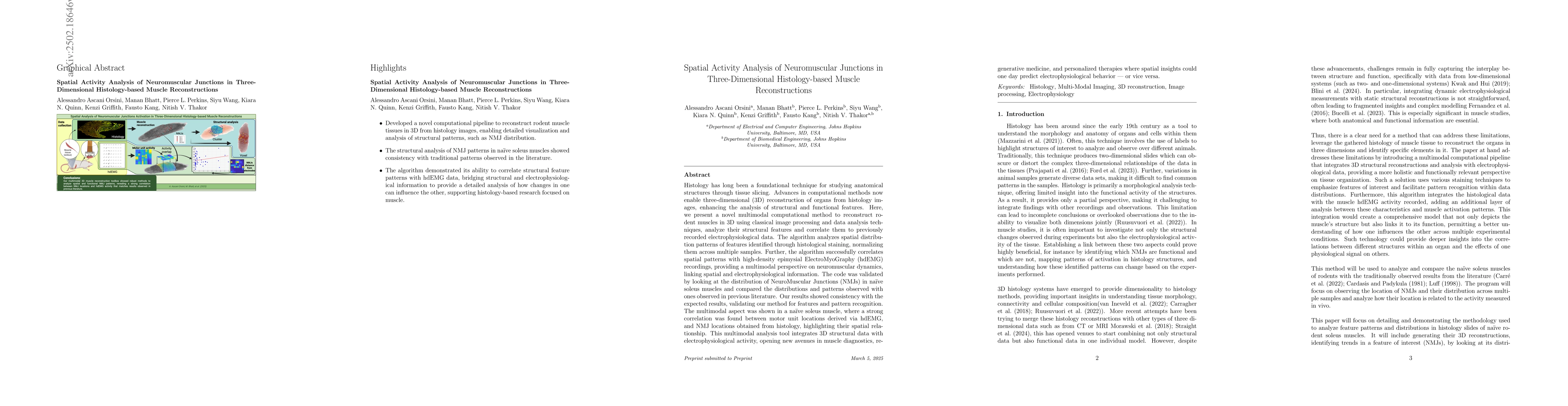
Histology has long been a foundational technique for studying anatomical structures through tissue slicing. Advances in computational methods now enable three dimensional (3D) reconstruction of organs from histology images, enhancing the analysis of structural and functional features. Here, we present a novel multimodal computational method to reconstruct rodent muscles in 3D using classical image processing and data analysis techniques, analyze their structural features and correlate them to previously recorded electrophysiological data. The algorithm analyzes spatial distribution patterns of features identified through histological staining, normalizing them across multiple samples. Further, the algorithm successfully correlates spatial patterns with high density epimysial ElectroMyoGraphy (hdEMG) recordings, providing a multimodal perspective on neuromuscular dynamics, linking spatial and electrophysiological information. The code was validated by looking at the distribution of NeuroMuscular Junctions (NMJs) in naive soleus muscles and compared the distributions and patterns observed with ones observed in previous literature. Our results showed consistency with the expected results, validating our method for features and pattern recognition. The multimodal aspect was shown in a naive soleus muscle, where a strong correlation was found between motor unit locations derived via hdEMG, and NMJ locations obtained from histology, highlighting their spatial relationship. This multimodal analysis tool integrates 3D structural data with electrophysiological activity, opening new avenues in muscle diagnostics, regenerative medicine, and personalized therapies where spatial insights could one day predict electrophysiological behavior or vice versa.

Key Findings, in focus

Seven facets of this paper, analysed and brought into focus by AI.

AI-generated · verify against source Generated Jun 11, 2025
In focus · one-line synthesis

This research integrates 3D structural data with electrophysiological activity, potentially opening new avenues in muscle diagnostics, regenerative medicine, and personalized therapies where spatial insights could predict electrophysiological behavior or vice versa.

01
MethodologyHow they did it

The research employs a novel multimodal computational method to reconstruct rodent muscles in 3D using classical image processing and data analysis techniques, analyzing their structural features and correlating them with electrophysiological data.

02
ResultsWhat they found
  • Validation of the method for feature and pattern recognition in NeuroMuscular Junction (NMJ) distribution in naive soleus muscles.
  • Strong correlation found between motor unit locations derived via high-density epimysial ElectroMyoGraphy (hdEMG) and NMJ locations obtained from histology, highlighting their spatial relationship.
03
SignificanceWhy it matters

This research integrates 3D structural data with electrophysiological activity, potentially opening new avenues in muscle diagnostics, regenerative medicine, and personalized therapies where spatial insights could predict electrophysiological behavior or vice versa.

04
Technical ContributionsWhat's new technically

The development of a novel computational method for 3D reconstruction and multimodal analysis of muscle structures, correlating them with electrophysiological data.

05
NoveltyWhat sets it apart

This work stands out by linking spatial and electrophysiological information in muscle analysis through 3D histology-based reconstructions, providing a comprehensive perspective on neuromuscular dynamics.

06
LimitationsWhat to be cautious about
  • The paper does not explicitly mention limitations, so this field is left blank based on the provided information.
07
Future WorkWhere it goes next
  • Further exploration of the method's applicability to other muscle types and species.
  • Investigating the predictive capabilities of spatial insights on electrophysiological behavior.

Impact

30
References

Paper Details

Paper ID: 2502.18646
Comments: 31 pages, 6 figures, for associated codes see: https://github.com/AleAsca/Histology-NMJ-and-muscle-Activity-Toolbox
Categories:
q-bio.QM

How to Cite This Paper

Reference manager: RIS EndNote

Citation Network

Current paper (gray), citations (green), references (blue)

Display is limited for performance on very large graphs.

Current Paper
Citations
References
Click to view


Discussion 0

This website uses cookies to ensure you get the best experience. Learn more