Summary
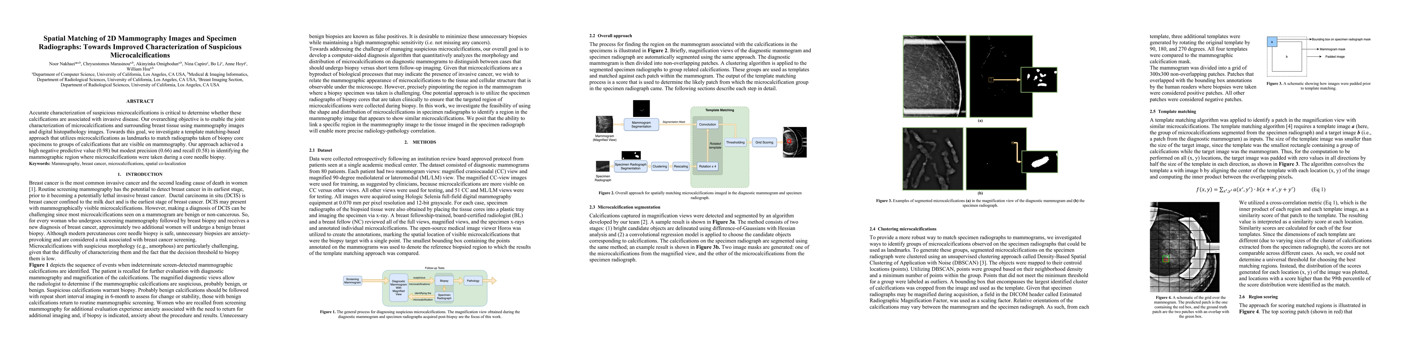
Accurate characterization of suspicious microcalcifications is critical to determine whether these calcifications are associated with invasive disease. Our overarching objective is to enable the joint characterization of microcalcifications and surrounding breast tissue using mammography images and digital histopathology images. Towards this goal, we investigate a template matching-based approach that utilizes microcalcifications as landmarks to match radiographs taken of biopsy core specimens to groups of calcifications that are visible on mammography. Our approach achieved a high negative predictive value (0.98) but modest precision (0.66) and recall (0.58) in identifying the mammographic region where microcalcifications were taken during a core needle biopsy.
AI Key Findings
Get AI-generated insights about this paper's methodology, results, and significance.
Paper Details
PDF Preview
Key Terms
Citation Network
Current paper (gray), citations (green), references (blue)
Display is limited for performance on very large graphs.
Similar Papers
Found 4 papersCost-effectiveness of contrast-enhanced breast MRI in suspicious mammographic microcalcifications.
Baltzer, Pascal A T, Fueger, Barbara J, Tollens, Fabian et al.
Findings of suspicious calcifications on contrast-enhanced mammography and their pathological correlation.
Mavili, Hanife Seda, Oral, Dilşah, Örgüç, İhsan Şebnem et al.
Mexican dataset of digital mammograms (MEXBreast) with suspicious clusters of microcalcifications.
Lozoya, Ricardo Salvador Luna, Barragán, Karina Núnez, Domínguez, Humberto de Jesús Ochoa et al.
No citations found for this paper.
Comments (0)