ArxivLens

Quick Summary:

This paper proposes a novel 3DCNN-based method for automated detection of multiple mitosis events in phase-contrast microscopy, addressing the limitations of existing methods by estimating a spatiotemporal likelihood map and mitigating annotation gaps. The method outperformed comparative methods in terms of F1-score on a challenging dataset.

arXiv Id: 2004.12531
Comments: 5 pages, 6 figures, Accepted in EMBC 2020
Date Published: 2020-06-02
Date Updated: 2020-06-02

Categories:

Key Terms (TF-IDF):

gaps (0.268)
gaps (0.267)
detect (0.219)
detection (0.218)
detection (0.209)
events (0.206)
events (0.203)
detect (0.200)
gap (0.188)
gap (0.187)
methods (0.169)
multiple (0.166)
gradually (0.165)
spatiotemporal (0.165)
proposed methods (0.165)
methods (0.164)
placed (0.162)
multiple (0.161)
annotated (0.161)
annotated (0.160)
License: http://arxiv.org/licenses/nonexclusive-distrib/1.0/

Summary:

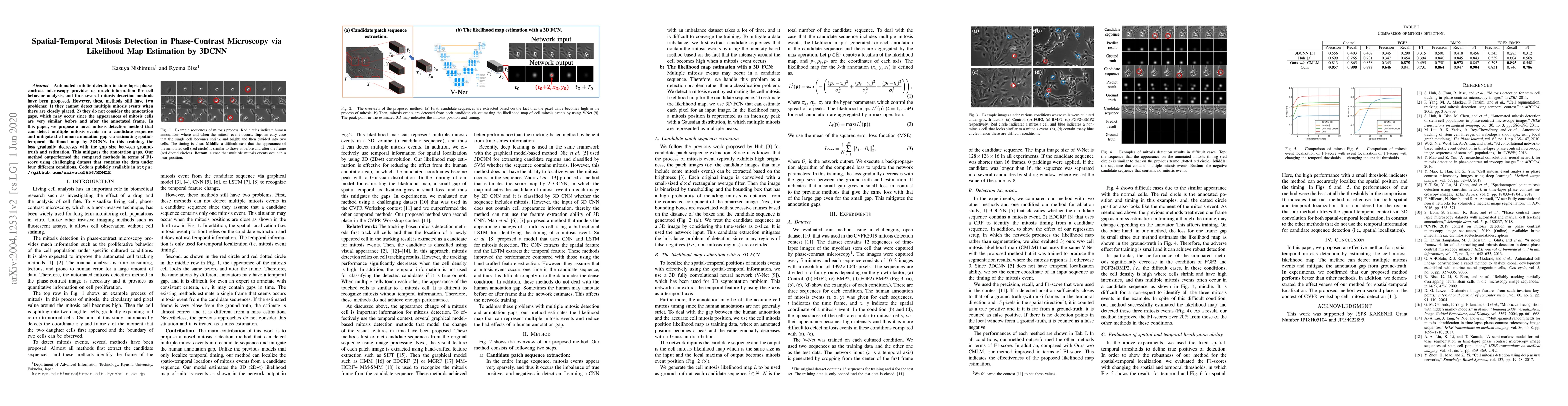
Automated mitotic detection in time-lapse phasecontrast microscopy provides us much information for cell behavior analysis, and thus several mitosis detection methods have been proposed. However, these methods still have two problems; 1) they cannot detect multiple mitosis events when there are closely placed. 2) they do not consider the annotation gaps, which may occur since the appearances of mitosis cells are very similar before and after the annotated frame. In this paper, we propose a novel mitosis detection method that can detect multiple mitosis events in a candidate sequence and mitigate the human annotation gap via estimating a spatiotemporal likelihood map by 3DCNN. In this training, the loss gradually decreases with the gap size between ground truth and estimation. This mitigates the annotation gaps. Our method outperformed the compared methods in terms of F1- score using a challenging dataset that contains the data under four different conditions.

Citation & Reference Network Visualization

This graph shows how this paper is connected to other research. The current paper is shown in dark gray, papers that cite it are in green, and papers it references are in blue. You can:

Current Paper
Papers citing this work (Citations)
Papers cited by this work (References)

• Hover over any node to see the paper title

• Click a node to view that paper's details

• Drag nodes to rearrange the graph

• Use the zoom controls (bottom right) to zoom in/out

Citations and References

Title Authors Year Actions
Automatic classification of normal and abnormal cell division using deep learning
Pablo Delgado-Rodriguez, Rodrigo Morales Sánchez, Elouan Rouméas-Noël, François Paris, A. Muñoz-Barrutia
2024
View Paper
Mitosis Detection from Partial Annotation by Dataset Generation via Frame-Order Flipping
Kazuya Nishimura, Ami Katanaya, S. Chuma, Ryoma Bise
2023
View Paper
DeepSea is an efficient deep-learning model for single-cell segmentation and tracking in time-lapse microscopy
Abolfazl Zargari, G. A. Lodewijk, N. Mashhadi, Nathan Cook, C. Neudorf, Kimiasadat Araghbidikashani, Robert Hays, Sayaka Kozuki, Stefany Rubio, E. Hrabeta-Robinson, Angela N. Brooks, L. Hinck, S. A. Shariati
2023
View Paper
Automatic Classification of Normal and Abnormal Cell Division Using Deep Learning Analysis of Mitosis Videos
Pablo Delgado-Rodriguez, Rodrigo Morales Sánchez, Elouan Rouméas-Noël, F. Paris, A. Muñoz-Barrutia
2023
View Paper
Mitosis Detection In Phase Contrast Microscopy Image Sequences Using Spatial Segmentation And Spatio-Temporal Localization Refinement
Ekansh Verma, V. Singh, M. Safwan
2021
View Paper

Comments (0)

This website uses cookies to ensure you get the best experience. Learn more