Spatially Multiplexed Interferometric Microscopy with one-dimensional diffraction grating
Publication
Metrics
AI Quick Summary
This paper reviews spatially multiplexed interferometric microscopy (SMIM) using a one-dimensional diffraction grating, demonstrating its application for super-resolved, reflective, and noise-reduced imaging, alongside single-shot amplitude and phase imaging. SMIM offers a cost-effective, simple, and stable method for converting standard microscopes into holographic ones.
Paper Preview
Abstract
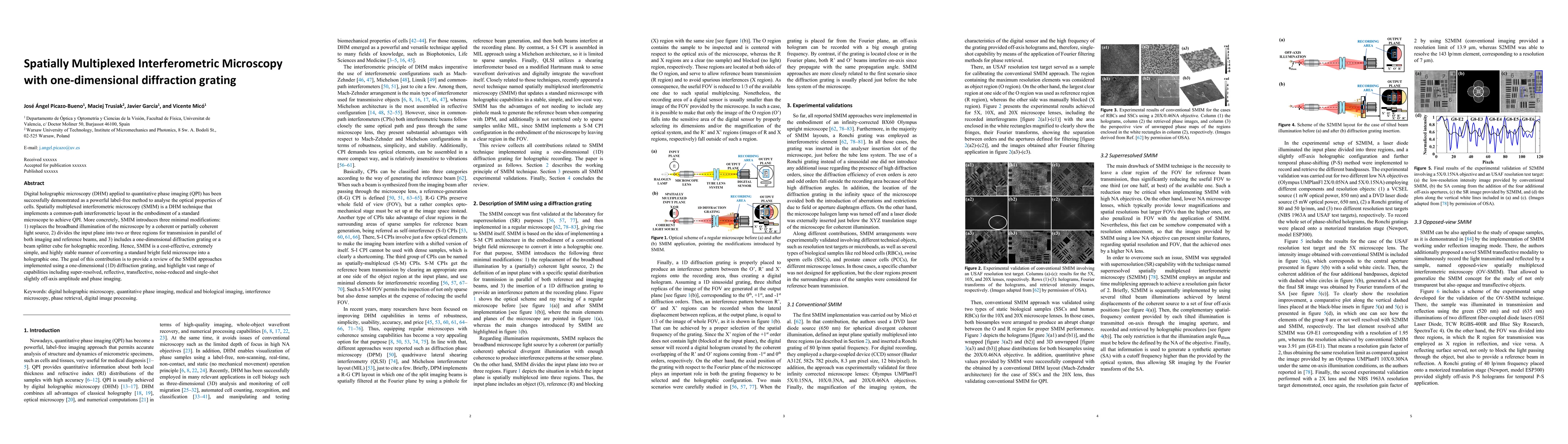
Digital holographic microscopy (DHM) applied to quantitative phase imaging (QPI) has been successfully demonstrated as a powerful label-free method to analyse the optical properties of cells. Spatially multiplexed interferometric microscopy (SMIM) is a DHM technique that implements a common-path interferometric layout in the embodiment of a standard microscope to achieve QPI. More concretely, SMIM introduces three minimal modifications: 1) replaces the broadband illumination of the microscope by a coherent or partially coherent light source, 2) divides the input plane into two or three regions for transmission in parallel of both imaging and reference beams, and 3) includes a one-dimensional diffraction grating or a beam splitter cube for holographic recording. Hence, SMIM is a cost-effective, extremely simple, and highly stable manner of converting a standard bright field microscope into a holographic one. The goal of this contribution is to provide a review of the SMIM approaches implemented using a one-dimensional (1D) diffraction grating, and highlight vast range of capabilities including super-resolved, reflective, transflective, noise-reduced and single-shot slightly off-axis amplitude and phase imaging.
AI Key Findings
Get AI-generated insights about this paper's methodology, results, significance, and more — seven facets brought into focus.
Authors
PDF Preview
Related Papers
No references found for this paper.
Discussion 0