This paper proposes a spatio-temporal multi-task learning approach for automatic quantification of cardiac left ventricle morphology from 3D Cine-MR images, aiming to reduce the time-consuming manual contouring process. The method segments the LV and predicts 11 indices and cardiac phases with high accuracy, demonstrating robustness across diverse cardiac morphologies.
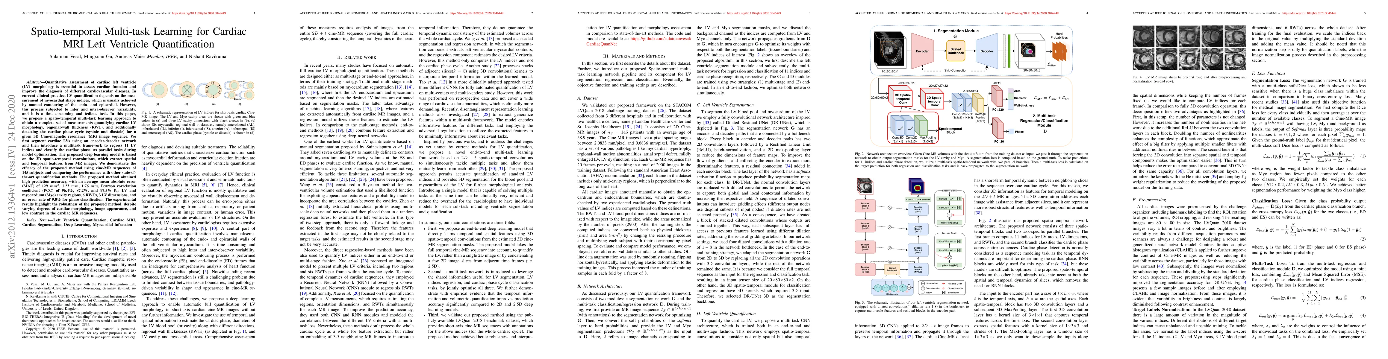
Quantitative assessment of cardiac left ventricle (LV) morphology is essential to assess cardiac function and improve the diagnosis of different cardiovascular diseases. In current clinical practice, LV quantification depends on the measurement of myocardial shape indices, which is usually achieved by manual contouring of the endo- and epicardial. However, this process subjected to inter and intra-observer variability, and it is a time-consuming and tedious task. In this paper, we propose a spatio-temporal multi-task learning approach to obtain a complete set of measurements quantifying cardiac LV morphology, regional-wall thickness (RWT), and additionally detecting the cardiac phase cycle (systole and diastole) for a given 3D Cine-magnetic resonance (MR) image sequence. We first segment cardiac LVs using an encoder-decoder network and then introduce a multitask framework to regress 11 LV indices and classify the cardiac phase, as parallel tasks during model optimization. The proposed deep learning model is based on the 3D spatio-temporal convolutions, which extract spatial and temporal features from MR images. We demonstrate the efficacy of the proposed method using cine-MR sequences of 145 subjects and comparing the performance with other state-of-the-art quantification methods. The proposed method obtained high prediction accuracy, with an average mean absolute error (MAE) of 129 $mm^2$, 1.23 $mm$, 1.76 $mm$, Pearson correlation coefficient (PCC) of 96.4%, 87.2%, and 97.5% for LV and myocardium (Myo) cavity regions, 6 RWTs, 3 LV dimensions, and an error rate of 9.0\% for phase classification. The experimental results highlight the robustness of the proposed method, despite varying degrees of cardiac morphology, image appearance, and low contrast in the cardiac MR sequences.
Get AI-generated insights about this paper's methodology, results, and significance.
Current paper (gray), citations (green), references (blue)
Title | Authors | Year | Actions |
---|---|---|---|
Multi-task Deep Learning in Medical Image Processing: A Systematic Review
|
J. Kamiri, Geoffrey Wambugu, A. Oirere
|
2025
|
View Paper |
MultiJSQ: Direct joint segmentation and quantification of left ventricle with deep multitask‐derived regression network
|
Xiuquan Du, Zheng Pei, Ying Liu, Xinzhi Cao, Lei Li, Shuo Li
|
2024
|
View Paper |
EFNet: A multitask deep learning network for simultaneous quantification of left ventricle structure and function.
|
Samana Batool, I. A. Taj, M. Ghafoor
|
2024
|
View Paper |
Simultaneous Deep Learning of Myocardium Segmentation and T2 Quantification for Acute Myocardial Infarction MRI
|
Yirong Zhou, Chengyan Wang, Mengtian Lu, Kunyuan Guo, Zi Wang, Dan Ruan, Rui Guo, Peijun Zhao, Jianhua Wang, Naiming Wu, Jianzhong Lin, Yinyin Chen, Hang Jin, Li-Ya Xie, Lilan Wu, Liuhong Zhu, Jianjun Zhou, Congbo Cai, He Wang, Xiaobo Qu
|
2024
|
View Paper |
Advancements in Left Ventricular Myocardium Analysis: A Comprehensive Review of Myocardium Segmentation, Fibrosis Detection and Wall Motion Estimation Using Cardiac Magnetic Resonance Imaging
|
Research Scholar, Dr. M. Kavitha
|
2024
|
View Paper |
Temporal-spatial Adaptation of Promptable SAM Enhance Accuracy and Generalizability of cine CMR Segmentation
|
Zhennong Chen, Sekeun Kim, Hui Ren, Quanzheng Li, Xiang Li
|
2024
|
View Paper |
Artificial Intelligence in Image-based Cardiovascular Disease Analysis: A Comprehensive Survey and Future Outlook
|
Xin Wang, Hongtu Zhu
|
2024
|
View Paper |
Data pyramid structure for optimizing EUS-based GISTs diagnosis in multi-center analysis with missing label
|
Lin Fan, Xun Gong, Cenyang Zheng, Jiao Li
|
2023
|
View Paper |
A task-unified network with transformer and spatial–temporal convolution for left ventricular quantification
|
Dapeng Li, Yanjun Peng, Jindong Sun, Yanfei Guo
|
2023
|
View Paper |
NVTrans‐UNet: Neighborhood vision transformer based U‐Net for multi‐modal cardiac MR image segmentation
|
Bingjie Li, Tiejun Yang, Xiang Zhao
|
2023
|
View Paper |
Multi-task deep learning for medical image computing and analysis: A review
|
Yan Zhao, Xiuying Wang, Tontong Che, Guoqing Bao, Shuyu Li
|
2022
|
View Paper |
MANET: Mitral Annulus Point Tracking Network in Cardiac Magnetic Resonance
|
Jianguo Chen, Xulei Yang, S. Leng, R. Tan, Zeng Zeng, L. Zhong
|
2022
|
View Paper |
Improving myocardial pathology segmentation with U-Net++ and EfficientSeg from multi-sequence cardiac magnetic resonance images
|
Hengfei Cui, Yan Li, Lei Jiang, Y. Wang, Yong Xia, Yanning Zhang
|
2022
|
View Paper |
Domain Generalization for Prostate Segmentation in Transrectal Ultrasound Images: A Multi-center Study
|
Sulaiman Vesal, Iani J. M. B. Gayo, Indrani Bhattacharya, S. Natarajan, Leonard S. Marks, D. Barratt, Richard E. Fan, Yipeng Hu, G. Sonn, M. Rusu
|
2022
|
View Paper |
Cardiac LGE MRI Segmentation With Cross-Modality Image Augmentation and Improved U-Net
|
Xinhua Yu, Junxin Chen, Bowen Fang, Wen Wang, Li-bo Zhang, Zhihan Lv
|
2021
|
View Paper |
Applying Cross-Modality Data Processing for Infarction Learning in Medical Internet of Things
|
Chenchu Xu, Zhifan Gao, D. Zhang, Jinglin Zhang, Lei Xu, Shuo Li
|
2021
|
View Paper |
Cardiac Magnetic Resonance 2D+T Short- and Long-axis Segmentation via Spatio-temporal SAM Adaptation
|
Zhennong Chen, Sekeun Kim, Hui Ren, Quanzheng Li, Xiang Li
|
2024
|
View Paper |
Comments (0)