Publication
Metrics
AI Quick Summary
This paper proposes a mathematical model to understand the spatio-temporal structure of Bone Multicellular Units (BMUs) in cortical bone, focusing on the interactions between osteoclasts and osteoblasts. The model reproduces experimentally-observed cell distributions and reveals how intrinsic parameters influence BMU dynamics, offering insights into bone physiology and disorders.
Paper Preview
Abstract
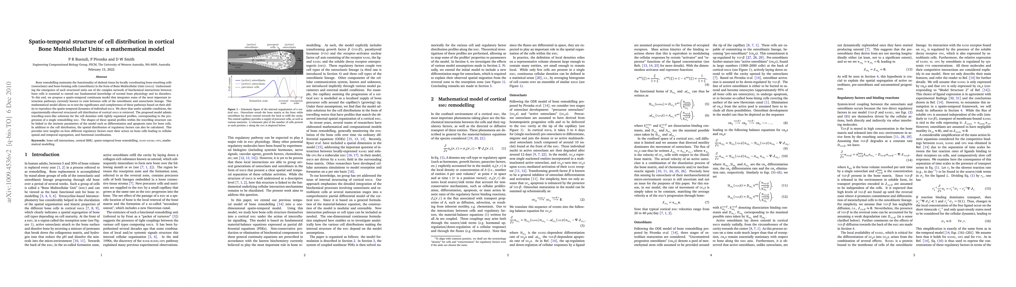
Bone remodelling maintains the functionality of skeletal tissue by locally coordinating bone-resorbing cells (osteoclasts) and bone-forming cells (osteoblasts) in the form of Bone Multicellular Units (BMUs). Understanding the emergence of such structured units out of the complex network of biochemical interactions between bone cells is essential to extend our fundamental knowledge of normal bone physiology and its disorders. To this end, we propose a spatio-temporal continuum model that integrates some of the most important interaction pathways currently known to exist between cells of the osteoblastic and osteoclastic lineage. This mathematical model allows us to test the significance and completeness of these pathways based on their ability to reproduce the spatio-temporal dynamics of individual BMUs. We show that under suitable conditions, the experimentally-observed structured cell distribution of cortical BMUs is retrieved. The proposed model admits travelling-wave-like solutions for the cell densities with tightly organised profiles, corresponding to the progression of a single remodelling BMU. The shapes of these spatial profiles within the travelling structure can be linked to the intrinsic parameters of the model such as differentiation and apoptosis rates for bone cells. In addition to the cell distribution, the spatial distribution of regulatory factors can also be calculated. This provides new insights on how different regulatory factors exert their action on bone cells leading to cellular spatial and temporal segregation, and functional coordination.
AI Key Findings
Get AI-generated insights about this paper's methodology, results, significance, and more — seven facets brought into focus.
Impact
Paper Details
PDF Preview
Key Terms
Citation Network
Current paper (gray), citations (green), references (blue)
Display is limited for performance on very large graphs.
Discussion 0