Authors
Summary
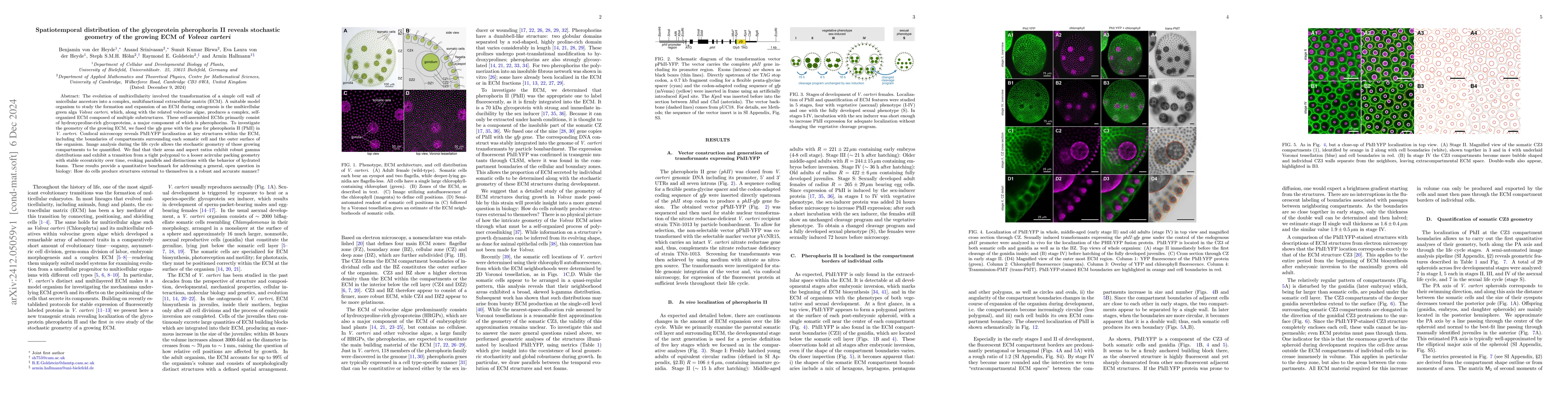
The evolution of multicellularity involved the transformation of a simple cell wall of unicellular ancestors into a complex, multifunctional extracellular matrix (ECM). A suitable model organism to study the formation and expansion of an ECM during ontogenesis is the multicellular green alga $Volvox~carteri$, which, along with the related volvocine algae, produces a complex, self-organized ECM composed of multiple substructures. These self-assembled ECMs primarily consist of hydroxyproline-rich glycoproteins, a major component of which is pherophorins. To investigate the geometry of the growing ECM, we fused the $yfp$ gene with the gene for pherophorin II (PhII) in $V.~carteri$. Confocal microscopy reveals PhII:YFP localization at key structures within the ECM, including the boundaries of compartments surrounding each somatic cell and the outer surface of the organism. Image analysis during the life cycle allows the stochastic geometry of those growing compartments to be quantified. We find that their areas and aspect ratios exhibit robust gamma distributions and exhibit a transition from a tight polygonal to a looser acircular packing geometry with stable eccentricity over time, evoking parallels and distinctions with the behavior of hydrated foams. These results provide a quantitative benchmark for addressing a general, open question in biology: How do cells produce structures external to themselves in a robust and accurate manner?
AI Key Findings
Generated Sep 02, 2025
Methodology
The study utilized a semi-automated image analysis pipeline with Cellpose, contrast stretching, J-invariant filtering, and user-prompted polygon inputs for segmentation and geometric analysis of Volvox carteri ECM structures. Affine transformations and whitening were applied to analyze domain properties.
Key Results
- Pherophorin II (PhII) localization at key ECM structures, including somatic cell boundaries and outer organism surface, was revealed using YFP fusion.
- Stochastic geometry of growing ECM compartments exhibited robust gamma distributions, transitioning from polygonal to acircular packing with stable eccentricity over time.
- ECM growth parallels and distinguishes from hydrated foam behavior, offering insights into cellular self-organization and robust structure formation.
Significance
This research provides a quantitative benchmark for understanding how cells produce external structures accurately and robustly, contributing to the broader field of biological self-organization and multicellularity.
Technical Contribution
Development of a semi-automated image analysis pipeline for studying the spatiotemporal distribution and geometry of extracellular matrices in Volvox carteri.
Novelty
This work uniquely combines genetic engineering (PhII-YFP fusion), advanced image analysis (Cellpose, J-invariant filtering), and quantitative geometric modeling to reveal the stochastic geometry of growing ECMs in Volvox carteri.
Limitations
- The study focused on Volvox carteri, which may limit the generalizability of findings to other multicellular organisms.
- Image analysis relied on specific markers (PhII-YFP), potentially overlooking other crucial ECM components.
Future Work
- Investigate ECM formation and dynamics in other multicellular organisms to validate findings and explore diversity.
- Explore additional ECM markers and components to gain a more comprehensive understanding of ECM structure and function.
Paper Details
PDF Preview
Citation Network
Current paper (gray), citations (green), references (blue)
Display is limited for performance on very large graphs.
No citations found for this paper.
Comments (0)